Yang Ji1,2, Borjan Gagoski 2,3, W. Scott Hoge 1,2, Yogesh Rathi 1,2, and Lipeng Ning 1,2
1Brigham and Women’s Hospital, Boston, MA, United States, 2Harvard Medical School, Boston, MA, United States, 3Boston Children’s Hospital, Boston, MA, United States
1Brigham and Women’s Hospital, Boston, MA, United States, 2Harvard Medical School, Boston, MA, United States, 3Boston Children’s Hospital, Boston, MA, United States
we propose a time-division multiplexing based echo-planar imaging (TDM-EPI) sequence, which can accelerate relaxation-diffusion MRI and standard dMRI by 2 or 3 folds.
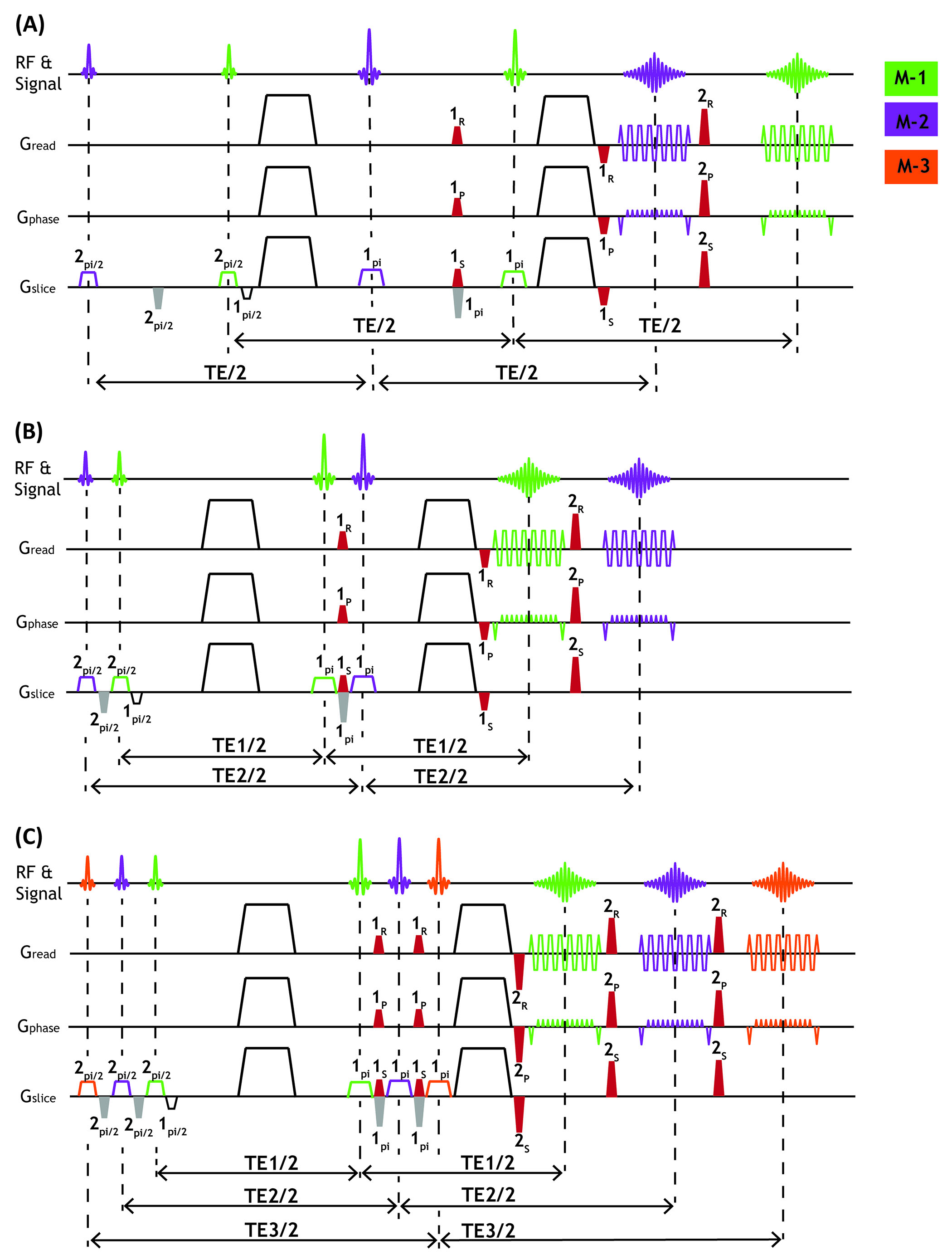
Figure1: Diagrams of the proposed
TDM-2s (B), TDM-2e (B) and TDM-3e EPI (C) sequences. The red gradient pulses
alternately dephase and rephase the echoes to separate the k-space of two TDM
slices, and gray gradients compensate for the phase dispersion induced by the
slice-selection gradients. Numbers listed on or below the gradients signify the
relative value of gradient area (i.e. zeroth moment) and corresponding subscripts
denote that gradients those have same subscripts share the same relative unit
standard.
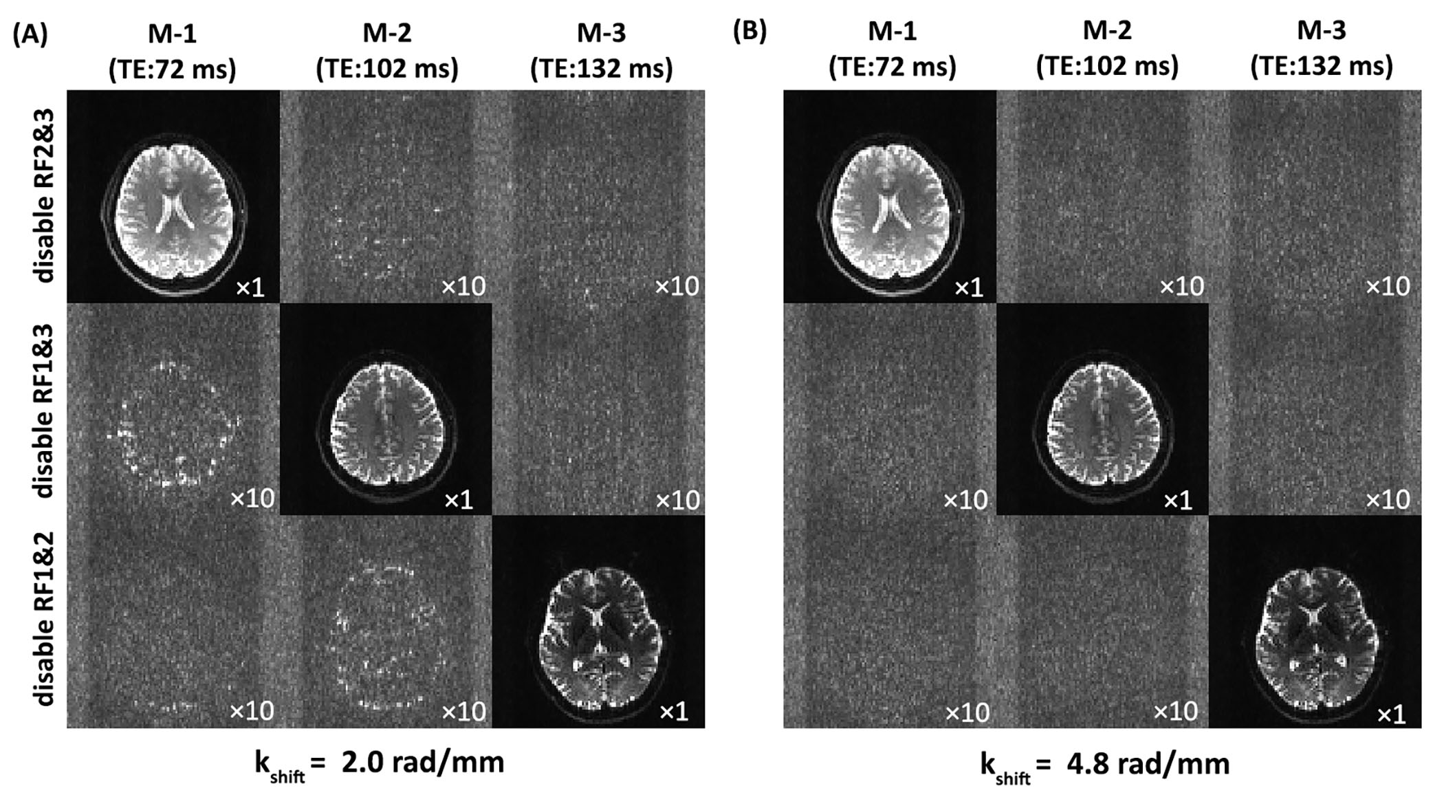
Figure
2:
Evaluation of signal leakage for
two representative shifting factors of 2.0 rad/mm (A) and 4.8 rad/mm (B) in
TDM-3e EPI sequence. To purely acquire the leaked signal of high spatial
frequencies from one slice to the others, only one pair of excitation and
refocusing RF pulses was enabled with the other two disabled in a single
sequence cycle. The numbers in the bottom right of the images represent the corresponding
enlargement factor.