Dinghui Wang1, Francis I. Baffour1, Daniel D. Borup2, Tzu-Cheng Chao1, and James G. Pipe1
1Radiology, Mayo Clinic, Rochester, MN, United States, 2Royal Philips, MR R&D, Rochester, MN, United States
1Radiology, Mayo Clinic, Rochester, MN, United States, 2Royal Philips, MR R&D, Rochester, MN, United States
A spiral dual echo spin-echo sequence with asymmetric in-out trajectories has been implemented and tested for simultaneous PDW and T2W knee imaging. Roughly a 20-36% gain in SNR can be achieved for the T2W images.
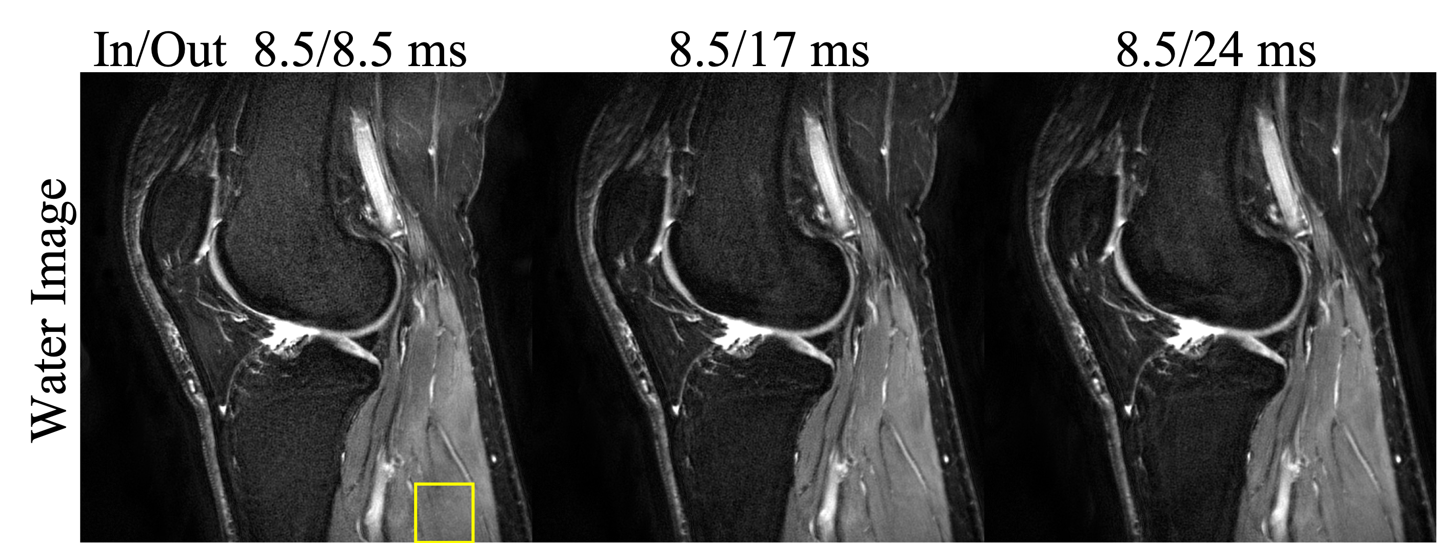
Figure 2 Comparison of spiral T2W water images of a right knee. Data were collected with difference spiral-in and spiral-out combinations. The ratio of SNR from left to right is 1: 1.22: 1.36 estimated inside the region shown by the yellow box.
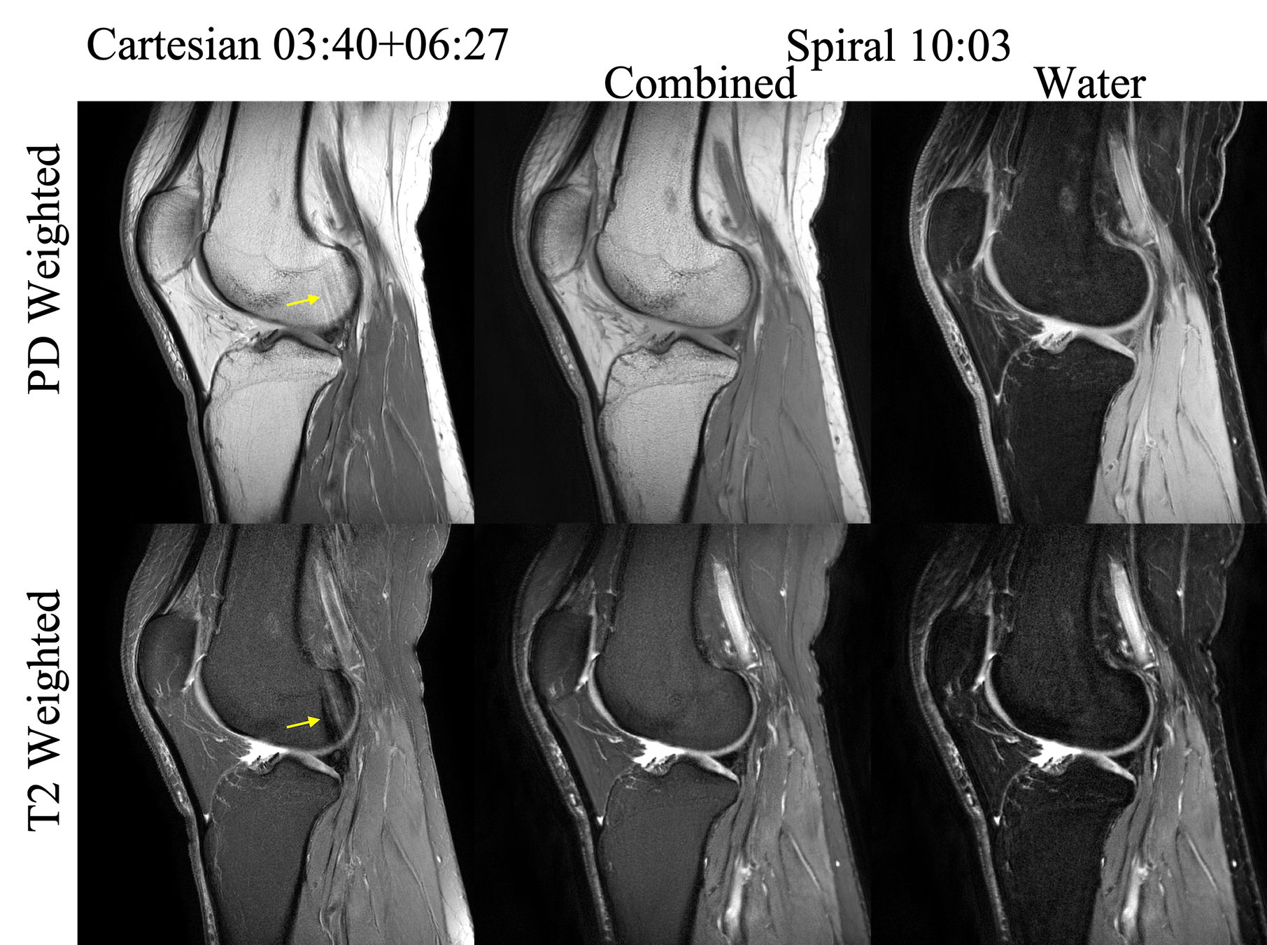
Figure 3 Sagittal PDW and T2W images of a right knee. Cartesian reference images, spiral water-fat combined images and spiral water only images are shown from left to right. In T2W spiral imaging, the fat only image (not shown) was partially (18%) combined with the water only image to form the water-fat combined image in the middle column. Spiral in/out lengths are 8.5/17ms. The yellow arrows point to flow artifacts seen in the Cartesian images, which are mitigated in the spiral images.