Jibin Tang1, Hongxi Zhang2, Zhipeng Shen3, Wenqi Wang1, Xingwang Yong1, Junjie Wen1, Xinchun Chen2, Fengyu Tian2, Weibo Chen4, Dan Wu1, and Yi Zhang1
1Key Laboratory for Biomedical Engineering of Ministry of Education, Department of Biomedical Engineering, College of Biomedical Engineering & Instrument Science, Zhejiang University, Hangzhou, Zhejiang, China, 2Department of Radiology, Children’s Hospital, Zhejiang University School of Medicine, Hangzhou, Zhejiang, China, 3Department of Neurosurgery, Children’s Hospital, Zhejiang University School of Medicine, Hangzhou, Zhejiang, China, 4Philips Healthcare, Shanghai, China
1Key Laboratory for Biomedical Engineering of Ministry of Education, Department of Biomedical Engineering, College of Biomedical Engineering & Instrument Science, Zhejiang University, Hangzhou, Zhejiang, China, 2Department of Radiology, Children’s Hospital, Zhejiang University School of Medicine, Hangzhou, Zhejiang, China, 3Department of Neurosurgery, Children’s Hospital, Zhejiang University School of Medicine, Hangzhou, Zhejiang, China, 4Philips Healthcare, Shanghai, China
We implemented a radiomic analysis of the APTw images, and found that the average sensitivity and AUC of the selected radiomic feature models for brain tumor grading were significantly higher than that of conventional mean APTw image intensities.
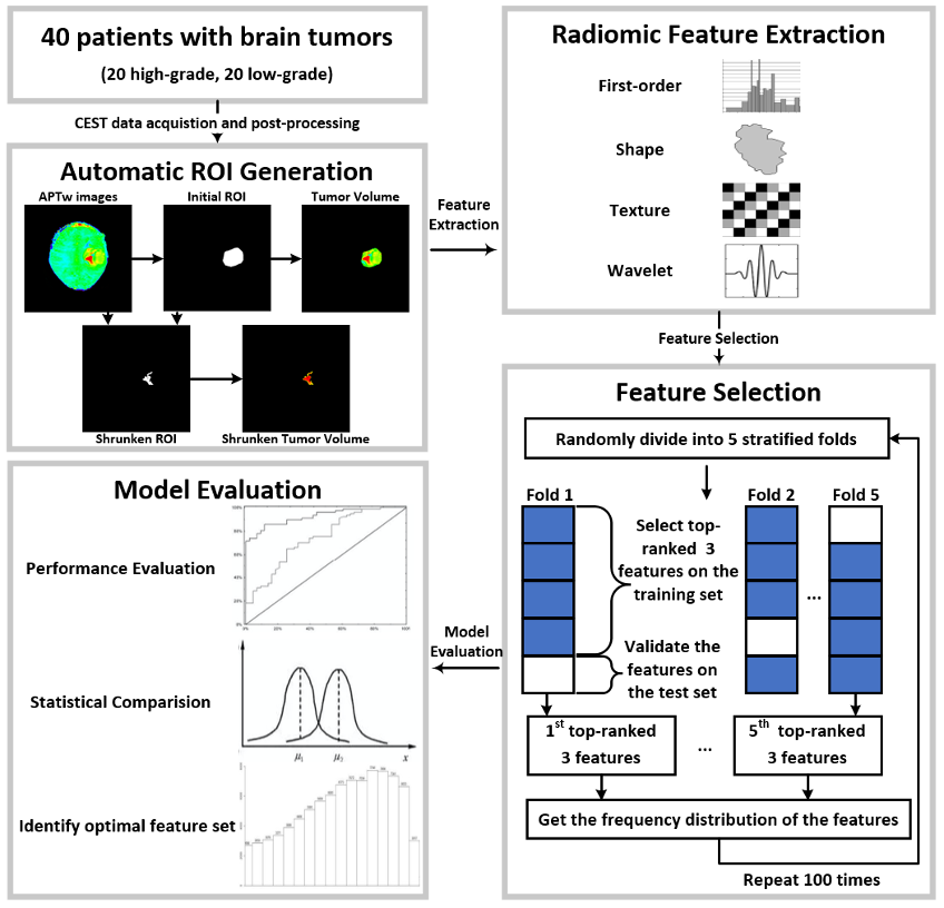
Figure 1. Flowchart of the whole process.
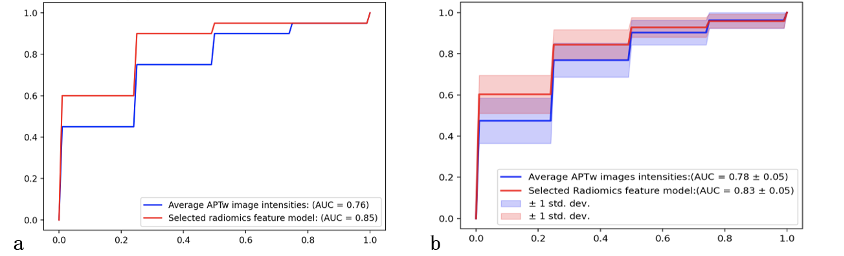
Figure 3. a: The average ROC curves of a representative 5-fold stratified cross-validation from radiomic analysis (red line) and coventional mean APTw signals (blue line). b: The pooled ROC curves (mean ± standard deviation) from 100 runs of the 5-fold stratified cross-validation, in which the red curves referred to results from radiomic analysis and blue curves denoted results from conventional mean APTw signals.