Henk M. De Feyter1, Monique A. Thomas1, Kevan L. Ip1, Kevin L. Behar2, and Robin A. de Graaf3,4
1Department of Radiology and Biomedical Imaging, Yale University, New Haven, CT, United States, 2Department of Psychiatry, Yale University, New Haven, CT, United States, 3Department of Radiology and Biomedical Imaging, Yale University, NEW HAVEN, CT, United States, 4Department of Biomedical Engineering, Yale University, New Haven, CT, United States
1Department of Radiology and Biomedical Imaging, Yale University, New Haven, CT, United States, 2Department of Psychiatry, Yale University, New Haven, CT, United States, 3Department of Radiology and Biomedical Imaging, Yale University, NEW HAVEN, CT, United States, 4Department of Biomedical Engineering, Yale University, New Haven, CT, United States
After in vivo
mapping of 2H-choline uptake with DMI, 2H NMR identifies
choline species in metabolite extracts from rat glioblastoma, acutely and 24 hrs
after infusion. The data show active metabolism of blood-borne choline in rat
glioblastoma, and long retention of 2H in choline metabolites.
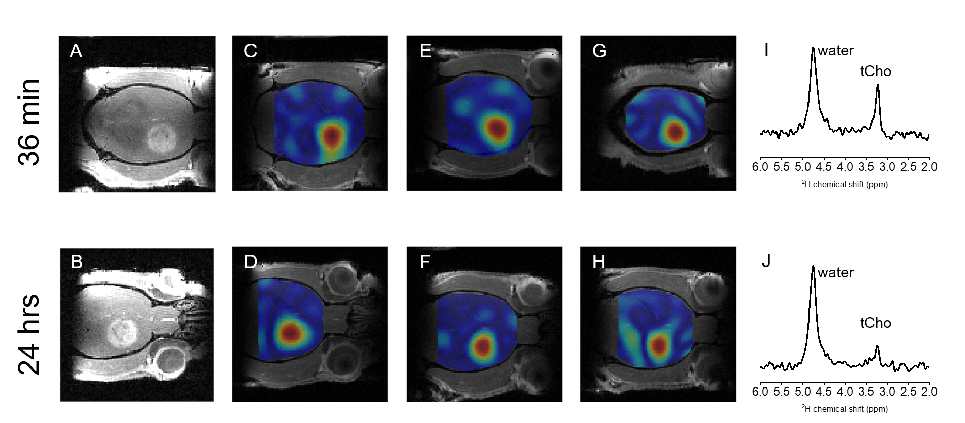
Figure
2. DMI of [2H9]-Cho in rats with RG2 glioma.
A,
B) Contrast-enhanced T1w MRI, C-G) DMI maps acquired during a ~36
min IV infusion of [2H9]-Cho, showing the high signal in
the tumor lesion in contrast to the surrounding normal brain. D-H), DMI maps
acquired 20-24 hrs after a ~36 min IV infusion of [2H9]-Cho;
note that panel A-D are from the same animal scanned on consecutive days. DMI
amplitude based on peak integral of tCho signal, in a.u., and normalized to the
highest value. I, J) 2H MR spectra from a voxel in the tumor,
acquired during and after 2H-choline infusion, respectively.
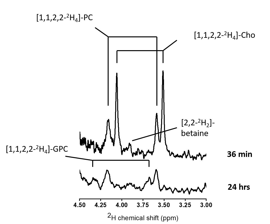
Figure
3. High resolution 2H NMR.
Spectra acquired in metabolite extracts from excised tumor tissue,
harvested immediately at the end (top), and 24 hrs after a 36 min infusion of
[1,1,2,2-2H4]-Cho (bottom), Note the lack of free Cho after
24 hrs, indicating active metabolism of the blood-borne 2H-labeled
Cho. Cho: choline, PC: phosphocholine, GPC, glycerophosphocholine. Note that
spectra are from samples of different weights, and thus peak amplitudes are not
necessarily quantitatively comparable.