Michael Wenke1, Jorg Dietrich2, Elizabeth Gerstner2, Otto Rapalino3, Julian He3, Daniel Kim1, Melanie Fu1, Pratik Talati4, Mohamed El Abtah1, Anna Vaynrub1, Sharif Natheir1, Mark Vangel3, Isabel Arrillaga-Romany2, Forst Deborah2, Yi-Fen Yen1, Ovidiu Andronesi1, Jayashree Kalpathy-Cramer1, Tracy Batchelor5, Bruce Rosen1, R. Gilberto Gonzalez3, and Eva-Maria Ratai1
1Radiology / Martinos Center for Biomedical Imaging, Massachusetts General Hospital, Charlestown, MA, United States, 2Neurology / Cancer Center, Massachusetts General Hospital, Boston, MA, United States, 3Radiology, Massachusetts General Hospital, Boston, MA, United States, 4Neurosurgery, Massachusetts General Hospital, Boston, MA, United States, 5Neurology, Brigham and Women's Hospital, Boston, MA, United States
1Radiology / Martinos Center for Biomedical Imaging, Massachusetts General Hospital, Charlestown, MA, United States, 2Neurology / Cancer Center, Massachusetts General Hospital, Boston, MA, United States, 3Radiology, Massachusetts General Hospital, Boston, MA, United States, 4Neurosurgery, Massachusetts General Hospital, Boston, MA, United States, 5Neurology, Brigham and Women's Hospital, Boston, MA, United States
Low tumoral myo-inositol prior to and during
anti-angiogenic therapy is predictive of poor survival.
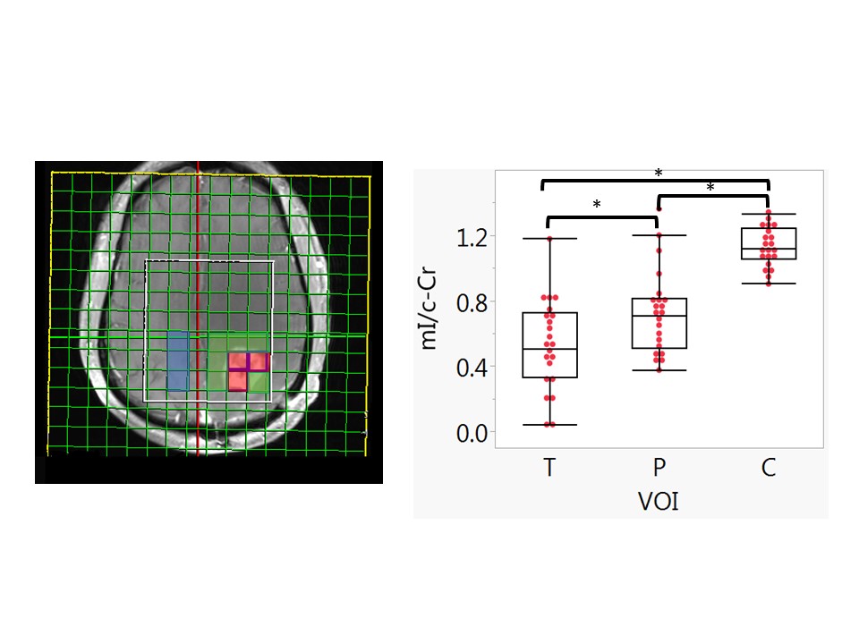
Figure 2. Left: Representative MRS voxel selection on a T1WI post-contrast image, tumor voxels (red), peritumoral (green), and contralateral normal voxels (blue) were selected for analyses.
Right: mI/c-Cr ratios at baseline in the tumor, (T) periphery (P), and contralateral (C) volumes of interest (VOI). Box plots indicate quartiles. Mean and standard deviations of mI/c-Cr in each VOI with t-tests indicating each VOI pair is significantly different.
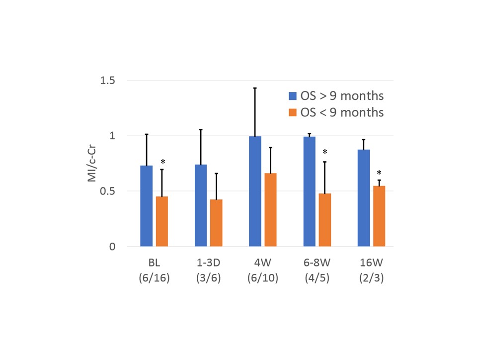
Figure
3. Mean and standard deviations of mI/c-Cr
classified by OS9. the asterisk (*) indicate p <0.05.