Georgios Batsios1, Celine Taglang1, Meryssa Tran1, Anne Marie Gillespie1, Joseph Costello2, Sabrina Ronen1, and Pavithra Viswanath1
1Radiology and Biomedical Imaging, UCSF, San Francisco, CA, United States, 2Neurological Surgery, UCSF, San Francisco, CA, United States
1Radiology and Biomedical Imaging, UCSF, San Francisco, CA, United States, 2Neurological Surgery, UCSF, San Francisco, CA, United States
Telomerase
reverse transcriptase (TERT) is essential for glioma proliferation and is an
attractive therapeutic target. Here, we show that TERT expression in gliomas is
linked to higher NADH, an effect that can be non-invasively monitored by
deuterium metabolic imaging using [U-2H]pyruvate.
Lactate
production from [U-2H]pyruvate is localized to the tumor region in vivo.
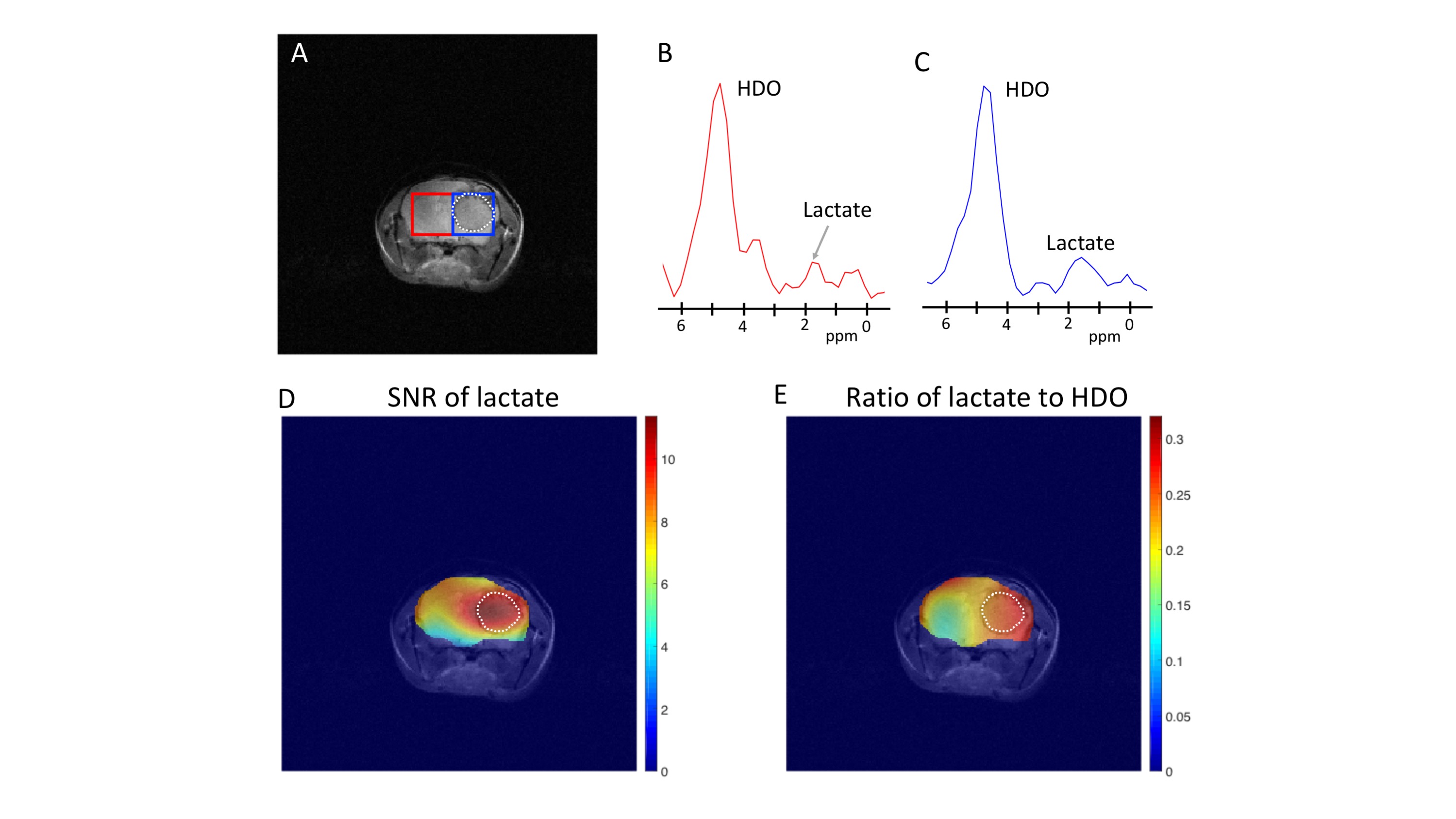
(A) Anatomical T2-weighted MRI of a
mouse bearing an orthotopic BT88 tumor xenograft. Representative 2H-MR spectra from
contralateral normal brain (B) and tumor (C) voxels at the first time
point after injection of [U-2H]pyruvate in a mouse bearing an orthotopic BT88 tumor. Metabolic heatmap of
the SNR of lactate (D) and the ratio
of lactate to post-injection HDO (E)
in mouse bearing an orthotopic BT88 tumor.
The tumor is delineated by white line.
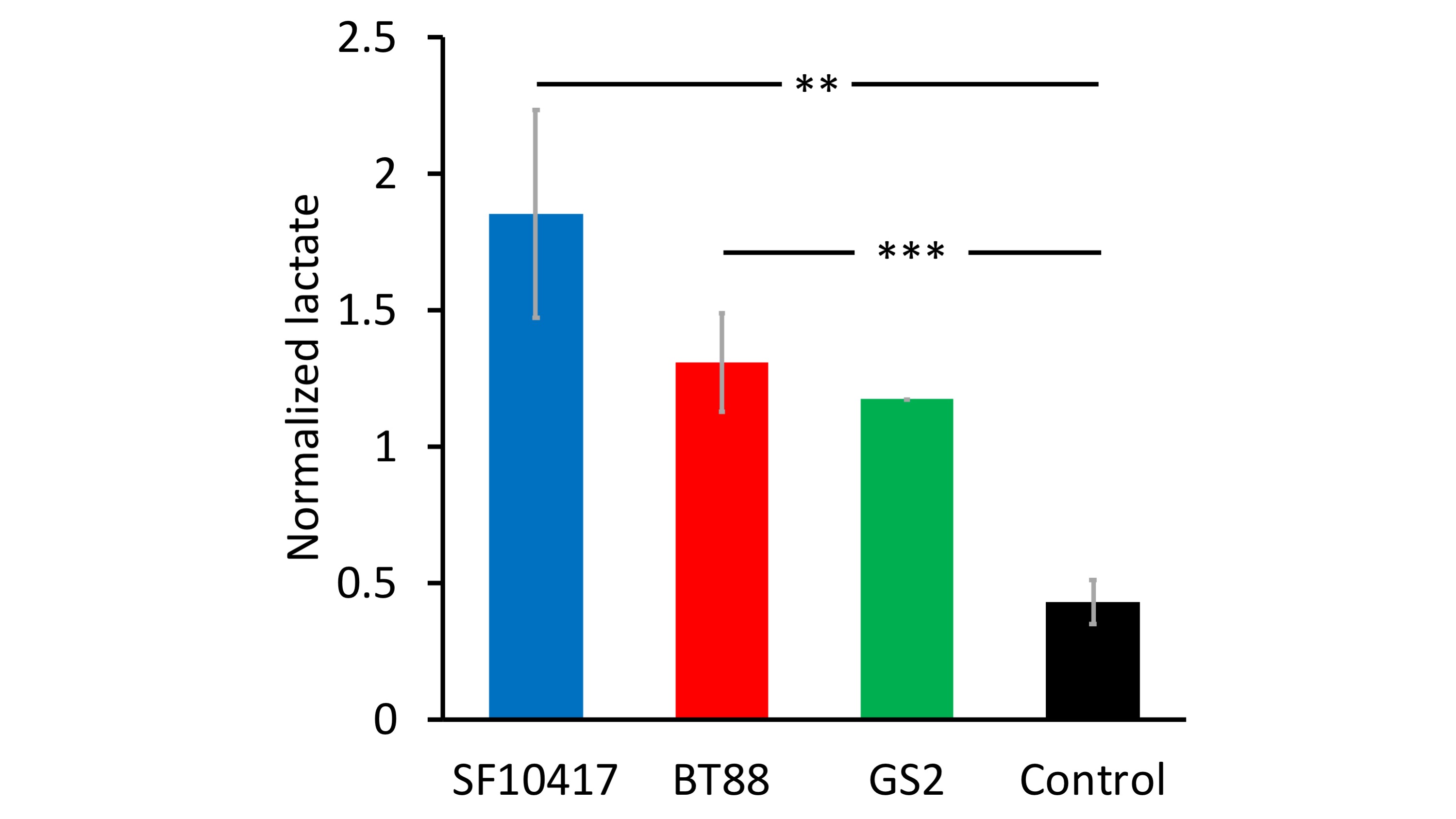
Lactate
production from [U-2H]pyruvate is higher in orthotopic glioma-bearing mice in
vivo. Lactate signal normalized to the ratio of post-injection HDO at
each time point to pre-injection HDO is higher in mice bearing orthotopic glioma
xenografts relative to tumor-free controls. ** refers to p<0.01, *** refers
to p<0.001.