Marina Radoul1, Donghyun Hong1, Anne Marie Gillespie1, Chloé Najac1, Pavithra Viswanath1, Russell O. Pieper2,3, Joseph Costello2, H. Artee Luchman4, and Sabrina M. Ronen1,3
1Radiology and Biomedical Imaging, University of California San Francisco, San Francisco, CA, United States, 2Neurological Surgery UCSF Weill Institute for Neurosciences, University of California San Francisco, San Francisco, CA, United States, 3Brain Tumor Research Center, University of California San Francisco, San Francisco, CA, United States, 4Hotchkiss Brain Institute, University of Calgary, Calgary, AB, Canada
1Radiology and Biomedical Imaging, University of California San Francisco, San Francisco, CA, United States, 2Neurological Surgery UCSF Weill Institute for Neurosciences, University of California San Francisco, San Francisco, CA, United States, 3Brain Tumor Research Center, University of California San Francisco, San Francisco, CA, United States, 4Hotchkiss Brain Institute, University of Calgary, Calgary, AB, Canada
Mutant IDH inhibition monitored in patient-derived glioma using in vivo 1H MRS revealed a decrease in 2HG and increase in Glu and GLX that were associated with subsequent slowdown of tumor growth and survival. This identifies early metabolic biomarkers of mutant IDH inhibition in glioma.

Figure 4: Representative axial T2-weighted images of control BT257 (A) and SF10417 (B) LGG-bearing mice and corresponding in vivo 1H MRS spectra (black) acquired from 2x2x2mm3 voxel marked in blue as well as the LCModel fit used to quantify the spectra (red) (A, B). Quantification of 2HG, Glu and GLX in control, AG-881- and BAY-1436032-treated tumor voxel at D7 and D15 in BT257 (C) and SF10417 (D) LGG-bearing mice.
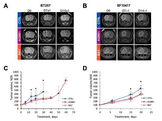
Figure 2: Representative axial T2-weighted images of control, AG-881 and BAY-1436032-treated BT257 (A) and SF10417 (B) LGG-bearing mice at D0, D7 and D15. Temporal evolution of average tumor volume shown as a percentage of D0 in BT257 (C) and SF10417 (D) LGG models.