Tanguy Boucneau1, Brice Fernandez2, Luc Darrasse1, and Xavier Maître1
1Université Paris-Saclay, CEA, CNRS, Inserm, BioMaps, Orsay, France, 2Applications & Workflow, GE Healthcare, Orsay, France
1Université Paris-Saclay, CEA, CNRS, Inserm, BioMaps, Orsay, France, 2Applications & Workflow, GE Healthcare, Orsay, France
Magnitude contrast MR Elastography
was developed on the basis of a motion-sensitizing magnetization preparation
to subsequently make use of any type of imaging sequence, like UTE or ZTE, to
mechanically characterize tissues, otherwise inaccessible with standard MRE.
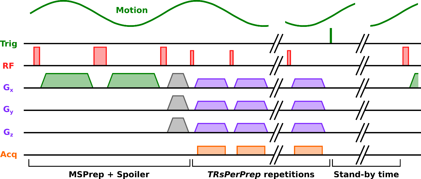
Figure 2: Representation of one segment of the UTE-based
MSPrep-MRE pulse sequence and delimitation of its three main parts: the motion
preparation and residual magnetization spoiling, the train of MRI repetitions
and the stand-by time (200 ms and 150 ms for HoP and HeP acquisitions
respectively).
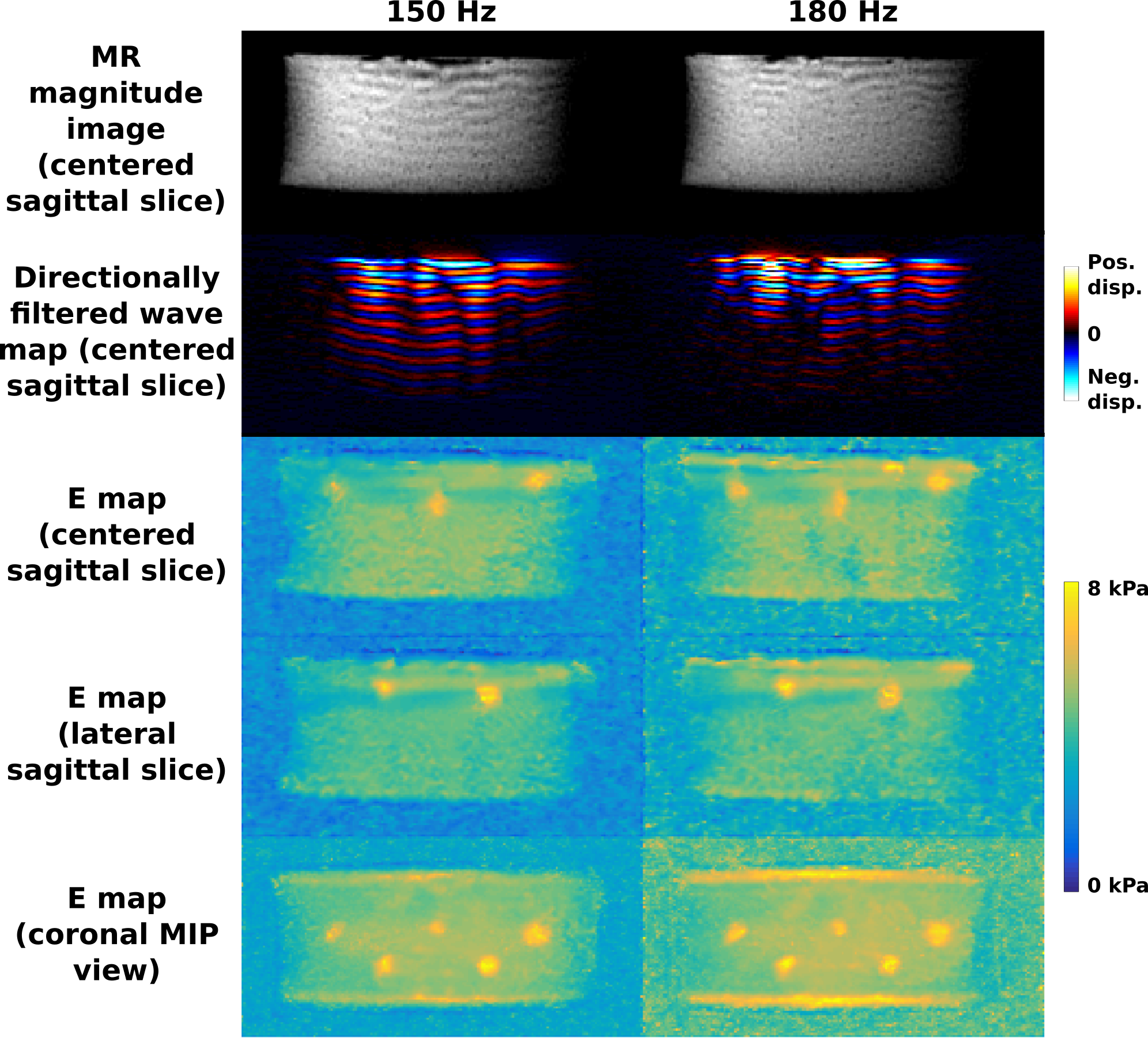
Figure 4: MR magnitude images, wave maps
directionally filtered along the up-to-down direction and Young’s modulus
maps obtained for the heterogeneous phantom (HeP) at 150 and 180 Hz. Two
sagittal planes with three and two inclusions are presented. A Maximum
Intensity Projection (MIP) in the coronal view clearly depicts the five
inclusions.