Martins Otikovs1, Noam Nissan2, Edna Furman-Haran1, Debbie Anaby2, Tanir M. Allweis3, Ravit Agassi4, Miri Sklair-Levy2,5, and Lucio Frydman1
1Weizmann Institute of Science, Rehovot, Israel, 2Sheba Medical Center, Ramat Gan, Israel, 3Kaplan Medical Center, Rehovot, Israel, 4Ben Gurion University Hospital, Beer Sheba, Israel, 5Tel Aviv University, Tel Aviv, Israel
1Weizmann Institute of Science, Rehovot, Israel, 2Sheba Medical Center, Ramat Gan, Israel, 3Kaplan Medical Center, Rehovot, Israel, 4Ben Gurion University Hospital, Beer Sheba, Israel, 5Tel Aviv University, Tel Aviv, Israel
Spatiotemporal encoding (SPEN) is an alternative ultrafast imaging technique. Its performance for breast cancer characterization using DW and diffusion kurtosis imaging and the potential of using SPEN DW images acquired with high b-values as an alternative to DCE subtractions are assessed.
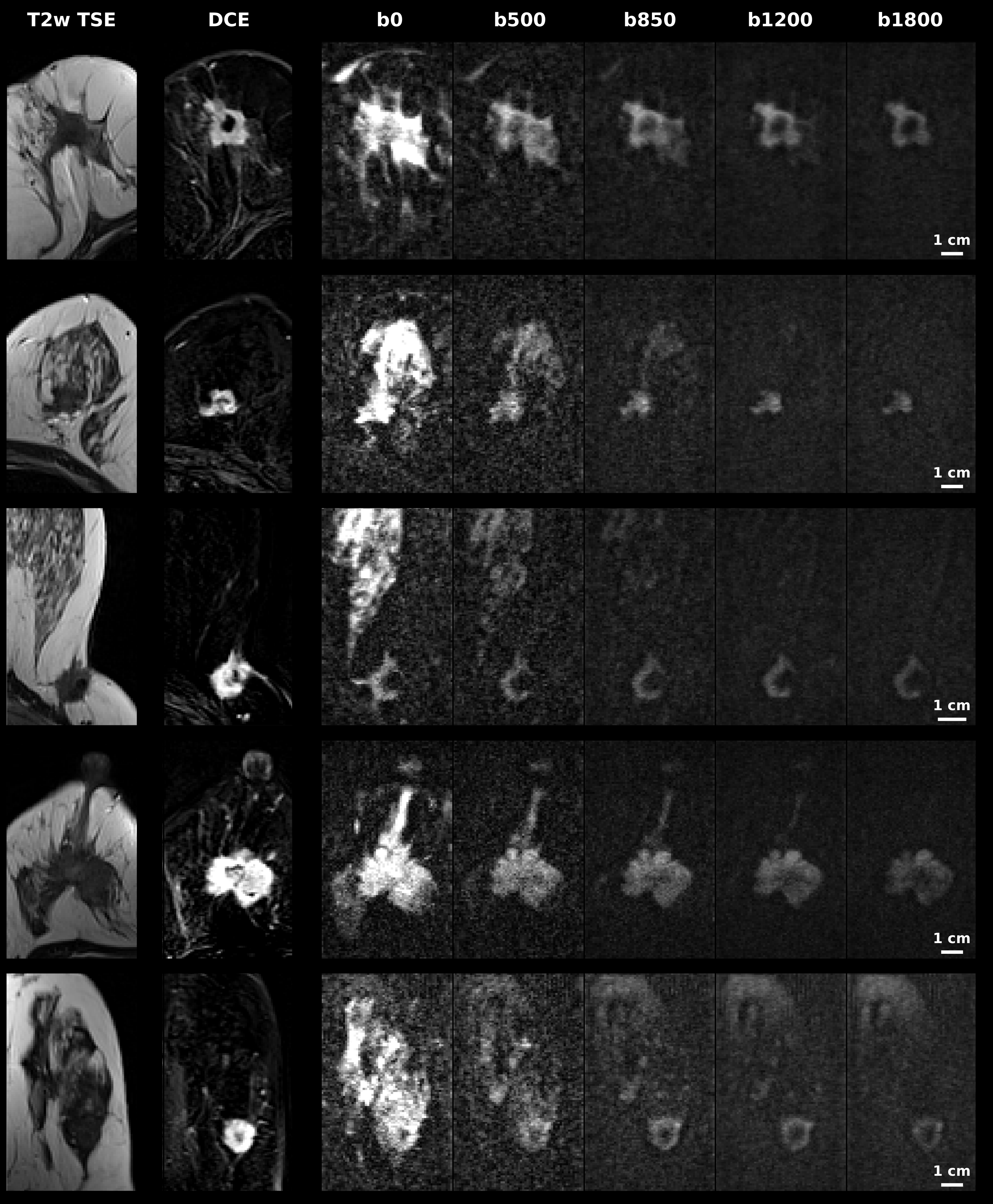
Figure 4. Comparison between DW images acquired using a series of b-encoding gradients (right-hand panels), and DCE subtraction images on the same patient. Diffusion encoding b-values are indicated on top of each column for SPEN DW images. In the two left columns T2w TSE and T1w DCE subtraction images are also presented. Shown are exams for four IDC (rows 1, 4, 5 and 6) and two ILC (rows 2 and 3) patients. These images suggests the possibility to identify and delineate lesions solely on the basis of strongly b-weighted images –without a need for contrast and for DCE subtractions.
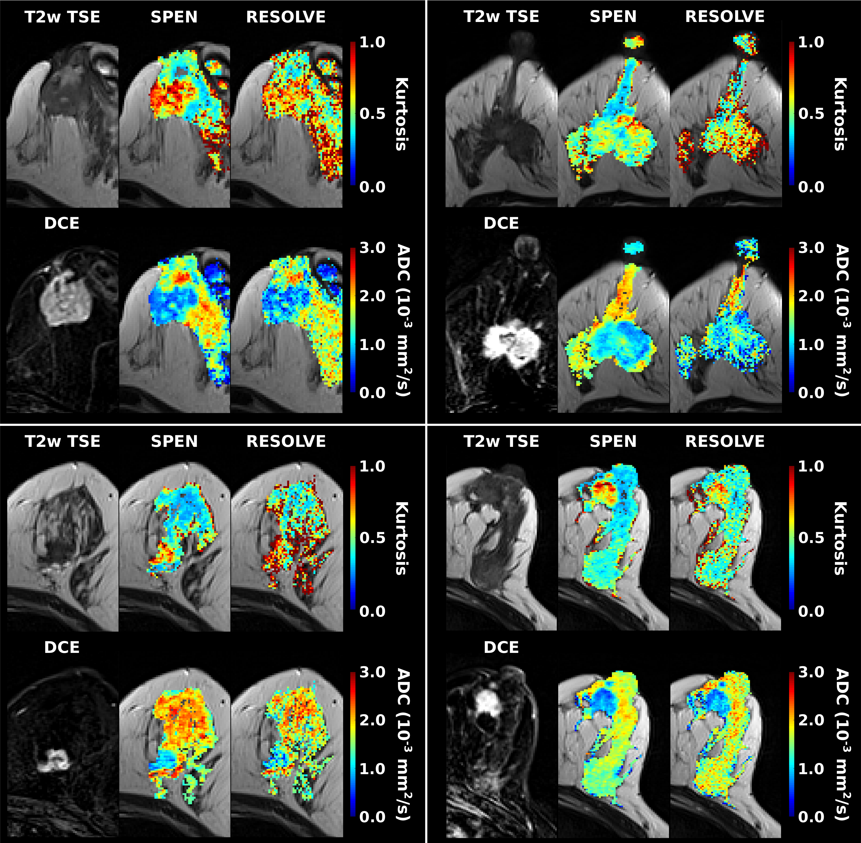
Figure 2. Representative ADC and kurtosis maps obtained from SPEN and RESOLVE data for four patients, displaying three IDC breast lesions and one ILC lesion (bottom left panel). Shown for each patient are single-breast anatomical T2w TSE and T1w DCE subtraction images, with the latter highlighting as bright regions the cancerous masses. Shown as well are the kurtosis and the ADC maps derived by the SPEN and RESOLVE exams (ADC maps were calculated solely on the basis of images acquired with 0 and 850 s/mm2 nominal b-values).