Jing Zhang1, Chenao Zhan2, Tao Ai2, Xu Yan3, and Guang Yang1
1Shanghai key lab of magnetic resonance, shanghai, China, 2Tongji Medical College, Huazhong University of Science and Technology, Department of Radiology,Tongji Hospital, Wuhan, Hubei Province, China, 3Siemens Healthcare, MR Scientific Marketing, shanghai, China
1Shanghai key lab of magnetic resonance, shanghai, China, 2Tongji Medical College, Huazhong University of Science and Technology, Department of Radiology,Tongji Hospital, Wuhan, Hubei Province, China, 3Siemens Healthcare, MR Scientific Marketing, shanghai, China
A
combined radiomics model using features from multi-parametric MRI achieved an
AUC of 0.948 to differentiate maglinant and benign breast cancers in the test
cohort, with an increased accuracy and a decreased false positive rate.
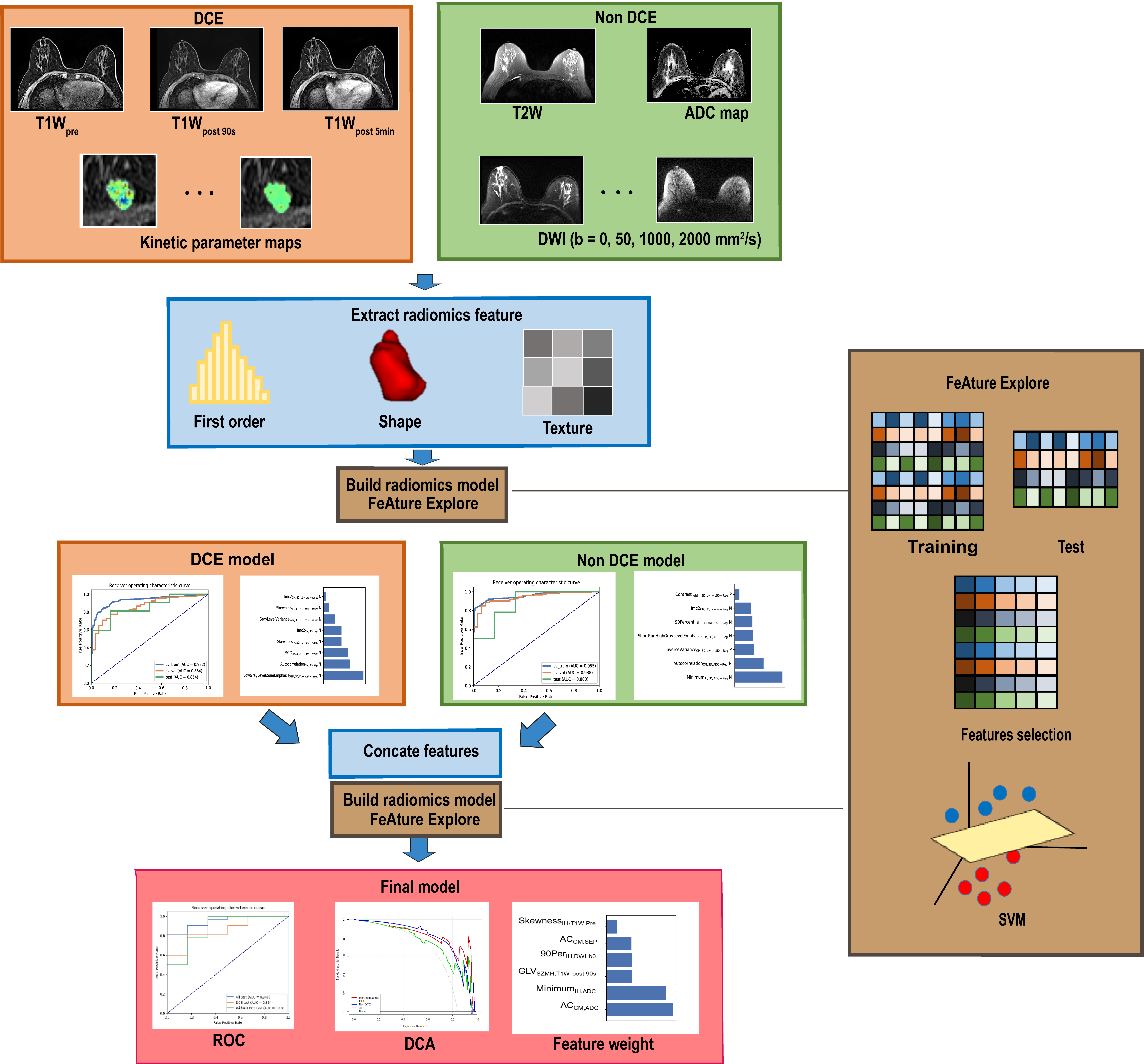
Figure 1 Flow chart. A tumor
region was contoured manually on T1Wpost 90s, to which other images
were aligned. First order and texture features were extracted from T1W,
DCE kinetic maps, T2W, and ADC maps, and used to build radiomics models,
together with shape features. The dataset was split into training and test cohort
by scanning date. After feature selection, radiomics models were constructed
using SVM or LR, before evaluated with ROC analysis, DCA
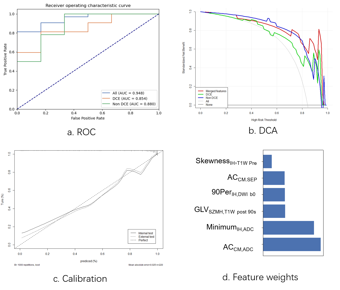
Figure
2 Model evaluation. (a) ROC in internel test
cohort. (b) Decision curve analysis for each model in the testing dataset. The
decision curve showed that when the threshold probability is in the range 0.85-0.95,
the application of the final model adds more benefit than treating all or none
of the patients and other models. (c) Calibration curve of the union model
prediction in the internal and external test cohort. (d) Weights of selected
features in the final model