Jean-Baptiste Perot1, Marina Célestine1, Marc Dhenain1, Sandrine Humbert2, Emmanuel Brouillet1, and Julien Flament1
1Université Paris-Saclay, Commissariat à l’Energie Atomique et aux Energies Alternatives (CEA), Centre National de la Recherche Scientifique (CNRS), Molecular Imaging Research Center (MIRCen), Laboratoire des Maladies Neurodégénératives, Fontenay-aux-Roses, France, 2Université Grenoble-Alpes, Grenoble Institute of Neurosciences (GIN), INSERM U1216, Grenoble, France
1Université Paris-Saclay, Commissariat à l’Energie Atomique et aux Energies Alternatives (CEA), Centre National de la Recherche Scientifique (CNRS), Molecular Imaging Research Center (MIRCen), Laboratoire des Maladies Neurodégénératives, Fontenay-aux-Roses, France, 2Université Grenoble-Alpes, Grenoble Institute of Neurosciences (GIN), INSERM U1216, Grenoble, France
Our longitudinal study including gluCEST, DTI, MT and rs-fMRI in a mouse model of Huntington's disease allowed detection of vulnerable brain networks. Early defects detected in white matter highlight its importance in the progression of the disease.
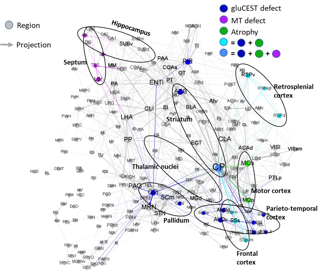
Figure 4: Summary of main results overlaid with axonal
projection atlas of the mouse brain. Grey arrows and dots represent axonal
projections and regions respectively. Colored dots represent affected regions
according to our results. Colored arrows represent axonal projections
between affected regions and can be associated with white matter alteration.
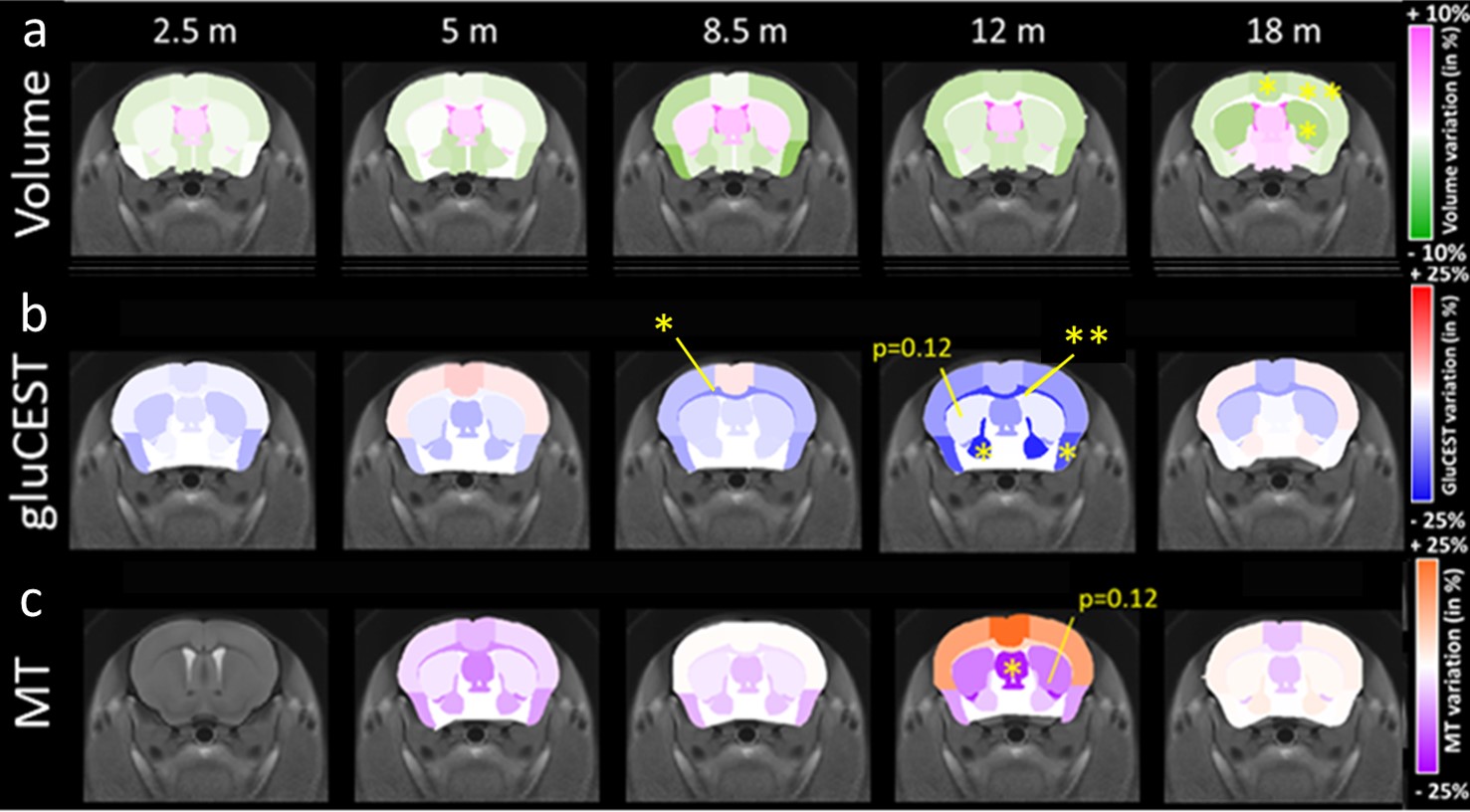
Figure 1: Variation
maps between Ki140 and WT mice. a) Volume variation maps, green regions show
atrophy in Ki140. b) GluCEST variation maps, blue regions show decreased level
in Ki140. c) MT variation maps, violet regions show decreased level in Ki140. *
p < 0.05, ** p<0.01.