Abdullah Althobity1,2, Nemat Khan3, Trent Woodruff3, Gary Cowin1,4, Ian Brereton1,4, and Nyoman Kurniawan1
1Centre for Advanced imaging, University of Queensland, Brisbane, Australia, 2Ministry of Education, Riyadh, Saudi Arabia, 3Faculty of Medicine, School of Biomedical Sciences, University of Queensland, Brisbane, Australia, 4National Imaging Facility, Brisbane, Australia
1Centre for Advanced imaging, University of Queensland, Brisbane, Australia, 2Ministry of Education, Riyadh, Saudi Arabia, 3Faculty of Medicine, School of Biomedical Sciences, University of Queensland, Brisbane, Australia, 4National Imaging Facility, Brisbane, Australia
MRS and DTI can detect pathological changes in EAE lumbar spinal
cord. Distinct metabolite and diffusion changes showed that complement protein
C5aR1 and C5L2 receptors play major roles in the progression of EAE.
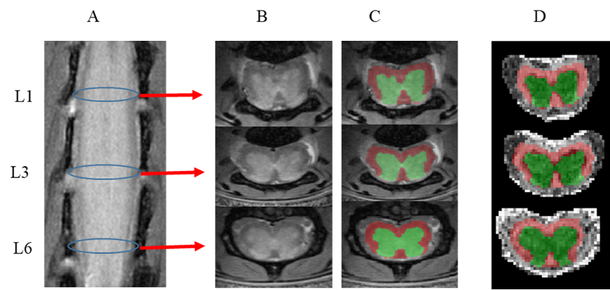
Figure 2. Anatomical and DTI analysis of the mouse lumbar spinal cord. (A) Coronal view showing the enlarged lumbar region. Blue circles indicate the positions for L1, L3 and L6. (B) High-resolution PD axial images. (C) GM/WM segmentation using SCT. (D) Affine registration of ROIs into DTI maps using b0. DTI parameters: number diffusion gradient directions = 9; b value = 1200 s/mm2 ; TR/TE = 800/18 ms; Resolution = 78 x 78 µm; Slice thickness = 0.5 mm; NSA = 1.
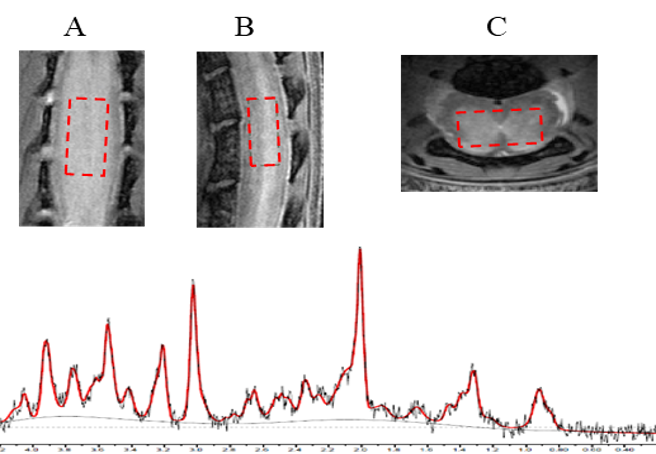
Figure 1. MRS of the lumbar spinal cord. High-resolution PD was used to locate enlarged lumbar spinal cord GM area. The dimension of MRS voxel is 2.8 x 1.6 x 0.9 mm as Z, X & Y directions as shown on A, B and C (coronal, sagittal and axial). PRESS was used with the following parameters: TR/TE = 2500/14 ms; number of signal averages (NSA) = 128; RF pulse band width = 8400Hz.