Paul Begovatz1, Lawrence Lechuga1, Monica Cho2, Mallery Olsen2, Rachel McMahon3, David Vail3,4, and Sean Fain1,5,6
1Medical Physics, Carbone Cancer Center, University of Wisconsin School of Medicine and Public Health, Madison, WI, United States, 2Pediatrics, Carbone Cancer Center, University of Wisconsin School of Medicine and Public Health, Madison, WI, United States, 3Medical Sciences, School of Veterinary Medicine, University of Wisconsin-Madison, Madison, WI, United States, 4Carbone Cancer Center, University of Wisconsin-Madison, Madison, WI, United States, 5Radiology, Carbone Cancer Center, University of Wisconsin School of Medicine and Public Health, Madison, WI, United States, 6Biomedical Engineering, Carbone Cancer Center, University of Wisconsin School of Medicine and Public Health, Madison, WI, United States
1Medical Physics, Carbone Cancer Center, University of Wisconsin School of Medicine and Public Health, Madison, WI, United States, 2Pediatrics, Carbone Cancer Center, University of Wisconsin School of Medicine and Public Health, Madison, WI, United States, 3Medical Sciences, School of Veterinary Medicine, University of Wisconsin-Madison, Madison, WI, United States, 4Carbone Cancer Center, University of Wisconsin-Madison, Madison, WI, United States, 5Radiology, Carbone Cancer Center, University of Wisconsin School of Medicine and Public Health, Madison, WI, United States, 6Biomedical Engineering, Carbone Cancer Center, University of Wisconsin School of Medicine and Public Health, Madison, WI, United States
This phantom feasibility study found that concentrations
of perfluoropolyether (PFPE), and fewer than one million PFPE labeled NK cells were
reliably detected through 19F-MRI with the combination of a cartesian
3D fast spin echo imaging sequence, and a dual tuned 1H/19F
torso coil at 3T.
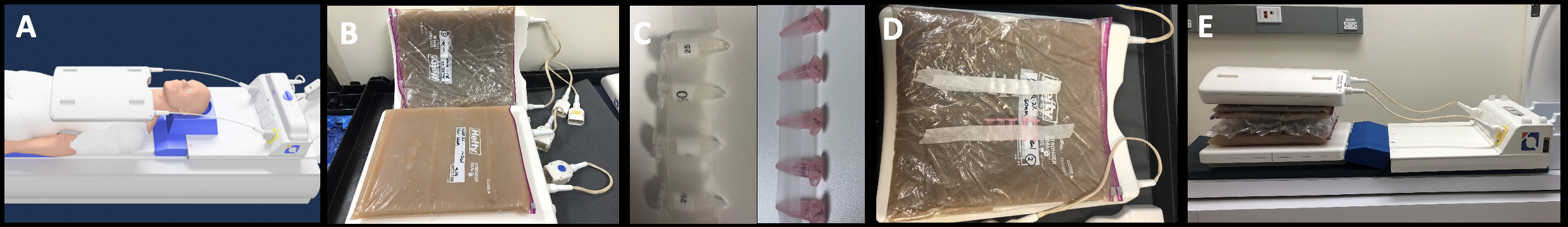
Figure
1. (A.) Dual tuned 1H/19F torso coil (MRI Tools, Berlin,
Germany), with (B.) agar spacer/loading phantoms, and (C.) 1.5 ml PFPE phantoms
(white) and 2 ml 19F labeled canine NK cell phantoms (pink). PFPE
phantoms were fixed to upper coil array elements under a single 4L agar loading
phantom approximately 2 cm from coil surface to simulate typical in vivo osteosarcoma
placement, and (D.) loaded in MRI with additional agar phantoms (4L, 8L) to
create spacing approximate to canine body.
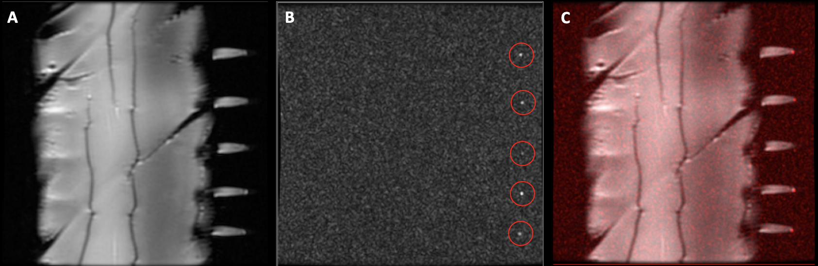
Figure 3. (A.) Coronal image of 2 cm thick 4L agar spacer and pelleted NK
cell phantoms. (B.) Typical 19F-image obtained from the cartesian 3D fast spin
echo sequence of the NK cell phantoms, with (C.) 19F-MRI overlay (red) of 1H-image
depicting the presence of 19F-signal within the tips of the pelleted NK cell
phantoms.