Dimitra Flouri1,2, Jack RT Darby3, Stacey L Holman3, Sunthara R Perumal4, Anna L David5,6, Janna L Morrison3, and Andrew Melbourne2,7
1School of Biomedical Engineering & Imaging Sciences, King's College London, London, United Kingdom, 2Department of Medical Physics & Biomedical Engineering, University College London, London, United Kingdom, 3Early Origins of Adult Health Research Group, University of South Australia, Adelaide, Australia, 4Preclinical Imaging and Research Laboratories, South Australian Health and Medical Research Institute, Adelaide, Australia, 5Elizabeth Garrett Anderson Institute for Women’s Health, University College London, London, United Kingdom, 6NIHR Biomedical Research Centre, University College London Hospitals, London, United Kingdom, 7School of Biomedical Engineering and Imaging Sciences, King's College London, London, United Kingdom
1School of Biomedical Engineering & Imaging Sciences, King's College London, London, United Kingdom, 2Department of Medical Physics & Biomedical Engineering, University College London, London, United Kingdom, 3Early Origins of Adult Health Research Group, University of South Australia, Adelaide, Australia, 4Preclinical Imaging and Research Laboratories, South Australian Health and Medical Research Institute, Adelaide, Australia, 5Elizabeth Garrett Anderson Institute for Women’s Health, University College London, London, United Kingdom, 6NIHR Biomedical Research Centre, University College London Hospitals, London, United Kingdom, 7School of Biomedical Engineering and Imaging Sciences, King's College London, London, United Kingdom
Here we characterised diffusion and perfusion properties of normal sheep placenta such as apparent diffusion coefficient, T2 measurements and fractional anisotropy analysis. We also presented the first application of multi-compartment MRI model to normal sheep placenta.
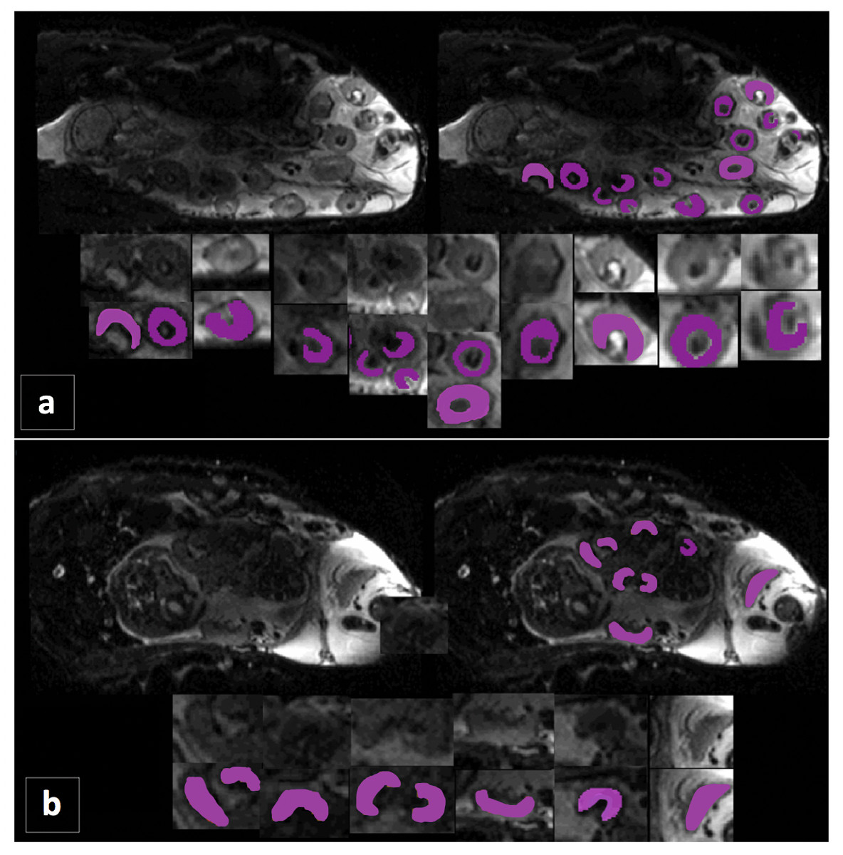
Figure 1: Example of MR images illustrating the placentomes from a single sheep at mid and late gestation.
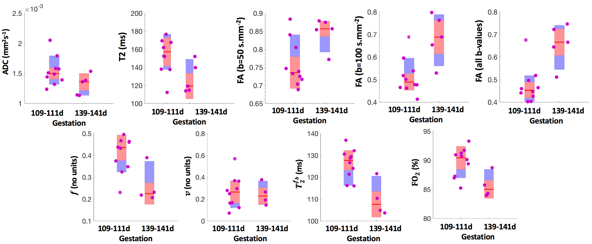
Figure 2: Boxplots summarising results over all singleton pregnancies at mid and late gestation. Each plot shows: the median (red line), the 25th and 75th percentile (purple box) and individual means of each sheep (pink circles).