Enrica Wilken1, Felix Freppon1, Max Masthoff1, and Cornelius Faber1
1Translational Research Imaging Center, Clinic of Radiology, University Hospital Muenster, Muenster, Germany
1Translational Research Imaging Center, Clinic of Radiology, University Hospital Muenster, Muenster, Germany
Repetitive T2*-weighted imaging of mice brain and simulations of velocity-dependent
blurring of contrast were used to show that single iron-labeled cells can be
resolved and tracked non-invasively by time-lapse MRI. Cell dynamics up to 1 µm/s
are detectable.
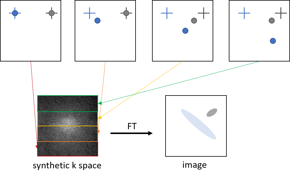
Figure 1: Principle of how image contrast of moving cells
with different velocities is simulated by creating synthetic k space.
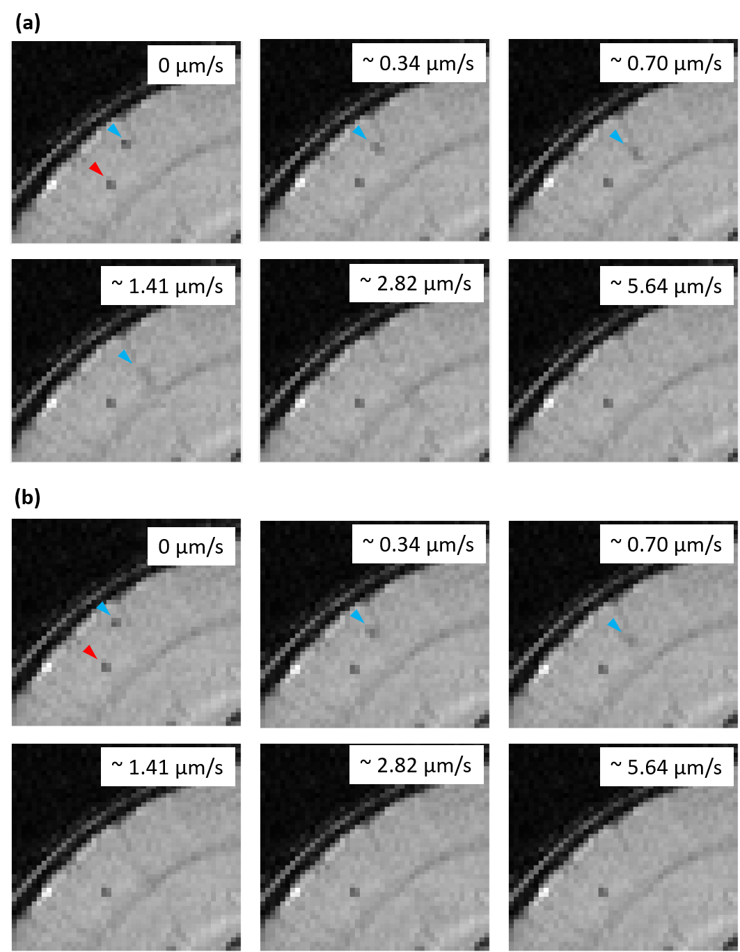
Figure 3: Image sections of an overlay of a T2*-weighted
image obtained by the time-lapse MRI protocol and the contrast simulation for
different cell motion velocities, showing a real (red arrowhead) and a
simulated (blue arrowhead) cell in the brain cortex. Simulations used a (a)
cartesian and (b) radial sampling scheme.