Maximilian Gram1,2, Markus Dippold2, Daniel Gensler1,3, Martin Blaimer4, Peter Nordbeck1,3, and Peter Michael Jakob2
1Department of Internal Medicine I, University Hospital Würzburg, Würzburg, Germany, 2Experimental Physics 5, University of Würzburg, Würzburg, Germany, 3Comprehensive Heart Failure Center (CHFC), University Hospital Würzburg, Würzburg, Germany, 4Fraunhofer Institute for Integrated Circuits IIS, Würzburg, Germany
1Department of Internal Medicine I, University Hospital Würzburg, Würzburg, Germany, 2Experimental Physics 5, University of Würzburg, Würzburg, Germany, 3Comprehensive Heart Failure Center (CHFC), University Hospital Würzburg, Würzburg, Germany, 4Fraunhofer Institute for Integrated Circuits IIS, Würzburg, Germany
Recently, a spin-lock-based technique for directly
measuring neuronal activity was introduced, which demonstrated imaging of alpha activity. In the present work it
is shown that the choice of the preparation parameters is of essential
importance and enables significant signal increases.

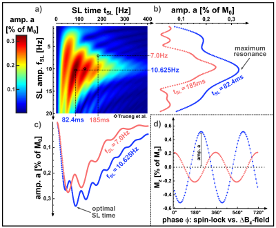
Figure 3) Simulation results of the parameter variation of tSL and fSL. The resulting NEMO-amplitude a is shown as a heat map (a). In (b) and (c) profiles for tSL and fSL are shown. In (d) the corresponding sinusoidal fits can be seen for varying phases ϕ. The optimal sequence parameters are tSL=82.4ms and fSL=10.625Hz and thus deviate significantly from the parameters chosen in [3] (125ms, 10Hz). In the simulation, typical relaxation times for gray matter T1ρ=78ms [3] and T2ρ=1.5*T1ρ were considered.

Figure 5) Results of the parameter variation. In (a) fSL was varied (constant tSL=100ms). Similar to the simulation, the NEMO-amplitude shows a peak at fSL≈10Hz. In (b) tSL was varied (constant fSL=10Hz). The emergence of local maxima can be seen. However, the behavior of RSS(tSL) is remarkable. In the case ≈25ms, ≈75ms and ≈125ms, image artifacts lead to great difficulties in the sinusoidal fit. A reliable determination of the NEMO-amplitude was therefore not possible in this range.
