Phan Tan Toi1,2,3, Hyun Jae Jang4, Jeehyun Kwag4, and Jang-Yeon Park1,2,3
1Department of Biomedical Engineering, Sungkyunkwan University, Suwon, Korea, Republic of, 2Center for Neuroscience Imaging Research, Institute for Basic Science, Suwon, Korea, Republic of, 3Department of Intelligent Precision Healthcare Convergence, Sungkyunkwan University, Suwon, Korea, Republic of, 4Department of Brain and Cognitive Engineering, Korea University, Seoul, Korea, Republic of
1Department of Biomedical Engineering, Sungkyunkwan University, Suwon, Korea, Republic of, 2Center for Neuroscience Imaging Research, Institute for Basic Science, Suwon, Korea, Republic of, 3Department of Intelligent Precision Healthcare Convergence, Sungkyunkwan University, Suwon, Korea, Republic of, 4Department of Brain and Cognitive Engineering, Korea University, Seoul, Korea, Republic of
In vivo direct MR imaging of neuronal activity at a high temporospatial
resolution
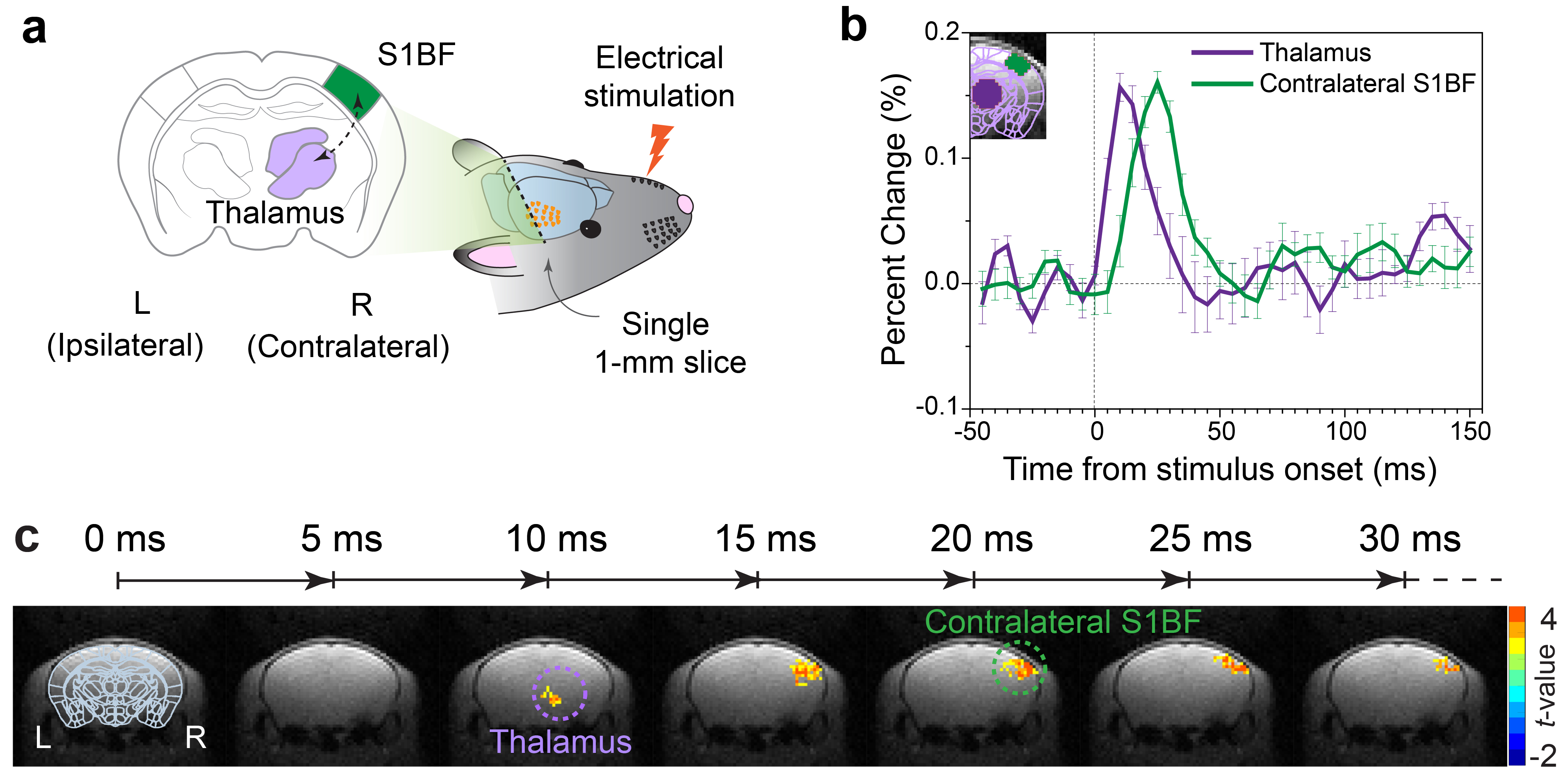
Figure 2. Temporospatial imaging of neuronal activity propagation. (a) Illustration of DIANA-fMRI experiment
with a coronal slice containing both thalamus and S1BF activated by electrical
stimulation applied to mouse whisker pad. (b) DIANA time series from
thalamus, contralateral S1BF (n = 10). Dotted line indicates the stimulation onset. (c) Time series of t-value maps for
30 ms after stimulation with 5 ms temporal resolution in one representative
mouse (paired t-test, p <
0.05, cluster size > 5 voxels). Dashed circular markers highlight the DIANA
response areas. Error bar: S.E.M.
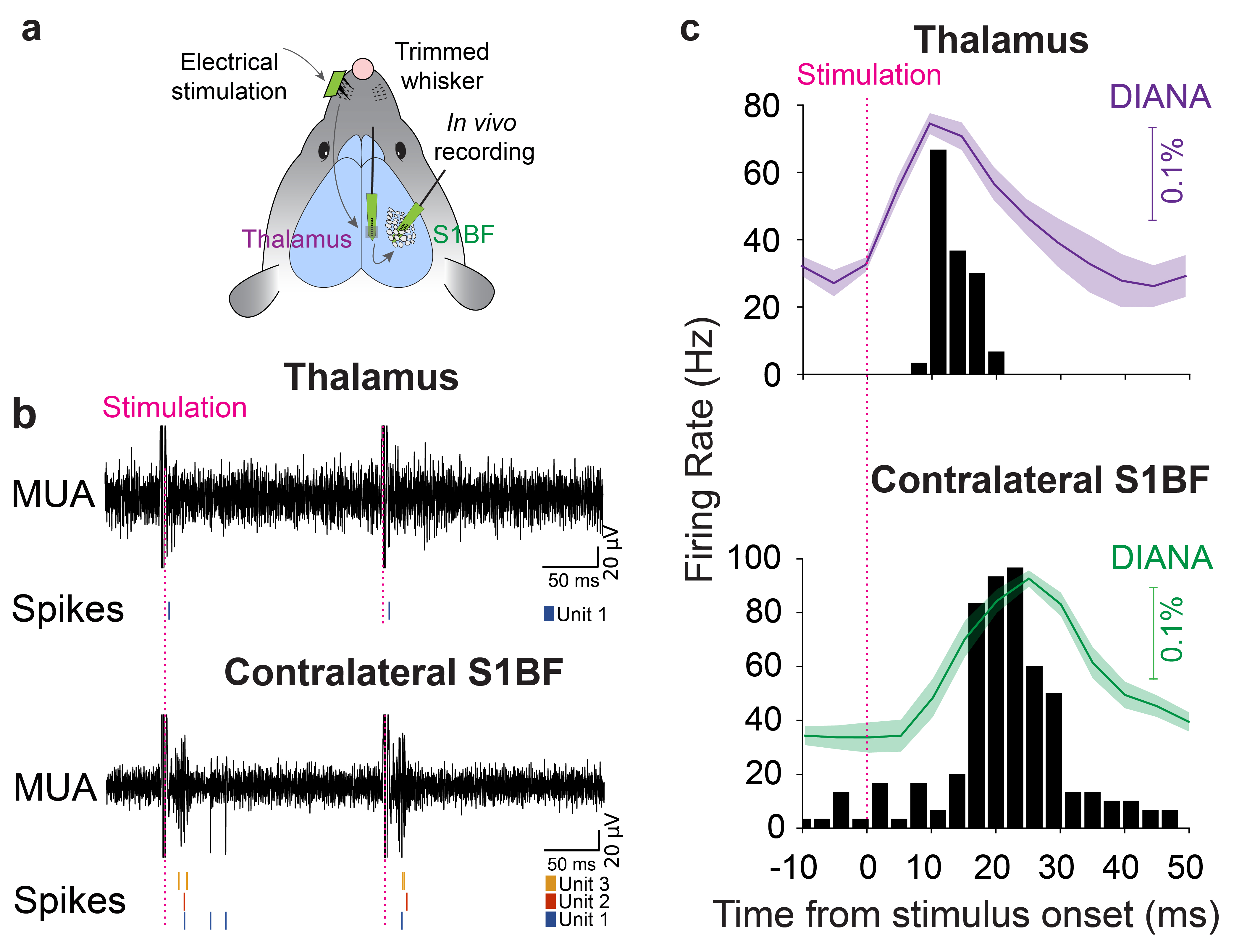
Figure 3. In vivo electrophysiological validation
of neuronal activity. (a)
Illustration of simultaneous electrophysiological recording in vivo both
thalamus and contralateral S1BF. (b) Representative MUA signals and
single-unit spikes acquired in thalamus (top) and contralateral S1BF (bottom).
(c) Post-stimulation time histogram of the responsive
single units over time plotted with DIANA signals in thalamus (top, 7 units from n = 7) and
contralateral S1BF (bottom, 16
units from n = 7). Dotted lines indicate the stimulation
onset. Shaded
area: S.E.M.
