Dan Benjamini1, Diego Iacono2, Michal Komlosh1, Daniel Perl2, David Brody2, and Peter Basser1
1National Institute of Child Health and Human Development, Bethesda, MD, United States, 2Uniformed Services University of the Health Sciences, Bethesda, MD, United States
1National Institute of Child Health and Human Development, Bethesda, MD, United States, 2Uniformed Services University of the Health Sciences, Bethesda, MD, United States
Can microscopic traumatic axonal injury be imaged noninvasively? We show that multidimensional MRI, which combines T1 , T2 and diffusion, can reveal damage that is invisible using quantitative MRI modalities, and provide an injury-only image that visualizes microscopic lesions in the brain.
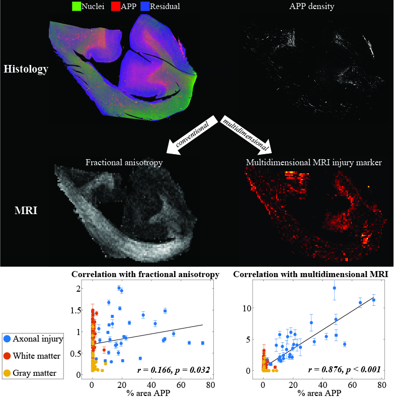
Summary of the findings, illustrated by comparing the FA with the T1-T2 injury image.
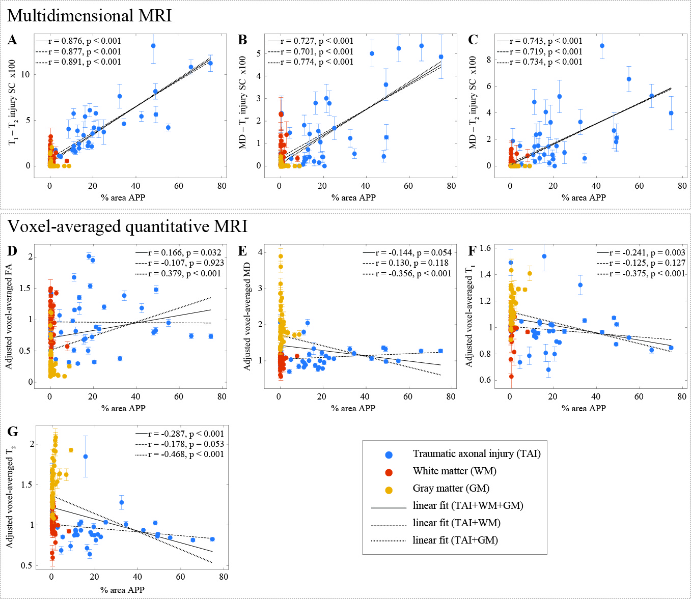
APP density (% area) from 132 tissue regions, consisting of 4 APP-positive regions from each TAI case (total of 32, blue dots), 4 to 6 normal-appearing WM regions from all cases (total of 56, red dots), and 4 cortical GM regions from all cases (total of 44, yellow dots), and the corresponding MR parameter correlations. Individual data points represent the mean ROI value from each post-mortem tissue sample. Scatterplots of the mean (with 95% confidence interval error bars) % area APP and all MR parameters.