Hyunyeol Lee1 and Felix W Wehrli1
1Radiology, University of Pennsylvania, Philadelphia, PA, United States
1Radiology, University of Pennsylvania, Philadelphia, PA, United States
Here, we develop a new whole-brain 3D quantitative BOLD parameter mapping method. The confounders (R2, non-heme iron) were addressed by employing prior estimates of R2’ and cerebral venous blood volume. Results suggest feasibility of the proposed, prior-based whole-brain 3D qBOLD mapping.
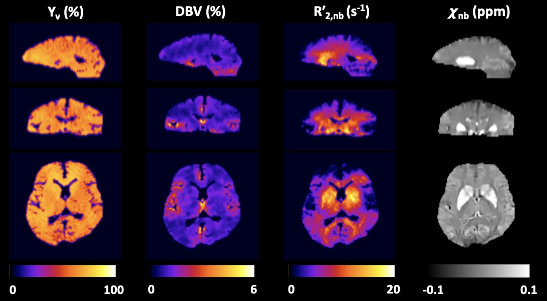
Figure 4. Whole-brain 3D maps of Yv, DBV,
R′2,nb, and χnb in
the three orthogonal planes, obtained using the proposed qBOLD method. Note
the expected contrast and physiologically plausible range in all four
parameters, i.e., a clear depiction of gray/white matter differentiation,
relatively homogeneous Yv, and elevated R′2,nb and χnb in
the deep brain structures relative to cortical regions.
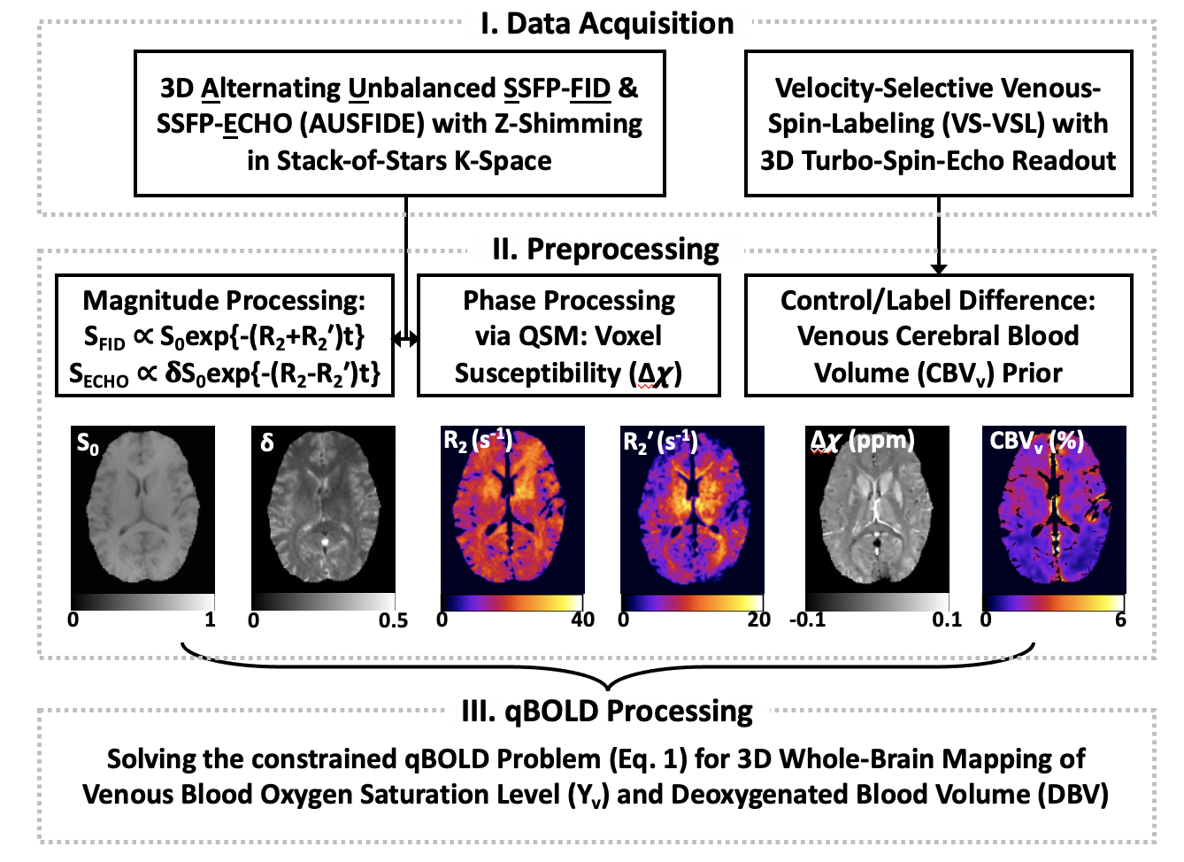
Figure 1. Schematic of the proposed,
preliminary estimates-based qBOLD parameter mapping procedure.