Sen Ma1, Tianle Cao1,2, Nan Wang1, Anthony G. Christodoulou1, Zhaoyang Fan1, Yibin Xie1, and Debiao Li1
1Biomedical Imaging Research Institute, Cedars-Sinai Medical Center, Los Angeles, CA, United States, 2Department of Bioengineering, University of California, Los Angeles, Los Angeles, CA, United States
1Biomedical Imaging Research Institute, Cedars-Sinai Medical Center, Los Angeles, CA, United States, 2Department of Bioengineering, University of California, Los Angeles, Los Angeles, CA, United States
We propose an integrated and efficient solution to clinical brain MRI in a single 10min sequence, producing co-registered, quantitative PD, T1, T2, T1ρ, T2*, QSM, and ΔB0 plus synthetic weighted images including PDw, T1w, T2w, T2*w, FLAIR, SWI, true-SWI, mIP, and true-SWI mIP simultaneously.
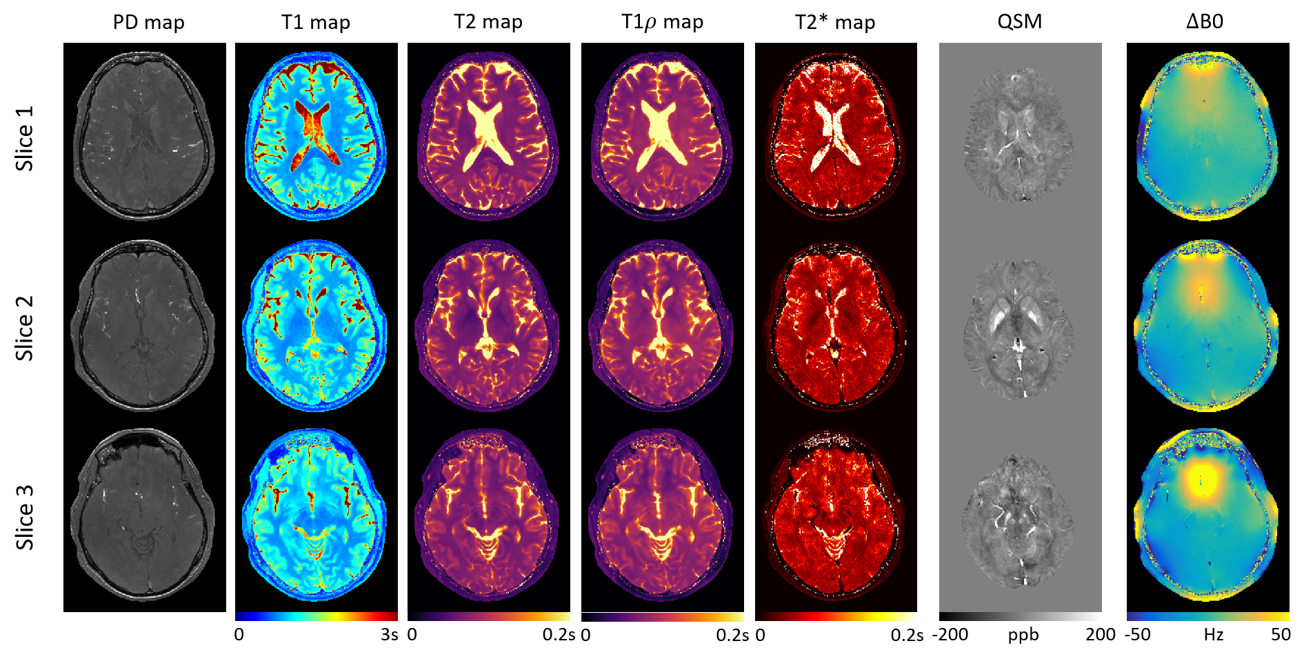
Figure 2. Axial PD, T1, T2, T1ρ,
T2*, QSM, and ∆B0
maps of three slices using the proposed
method.
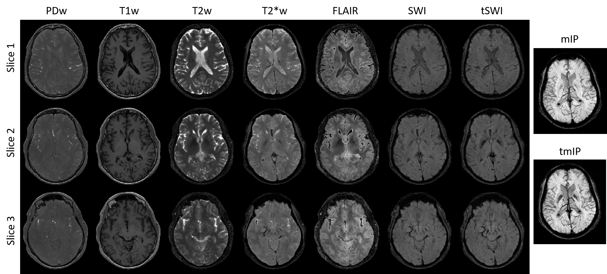
Figure 4. Synthetic PDw,
T1w, T2w, T2*w, FLAIR, SWI, tSWI of three axial slices
corresponding to Figure 2, as well as mIP and tmIP calculated from SWI and tSWI
respectively using 16 axial slices. Contrast-weighted images are synthesized
with good
image quality, image contrasts, and venous structures.