Sisi Li1, Yishi Wang2, Zhangxuan Hu3, Zhe Zhang4, Bing Wu3, and Hua Guo1
1Center for Biomedical Imaging Research, Beijing, China, 2Philips Healthcare, Beijing, China, 3GE Healthcare, MR Research China, Beijing, China, 4China National Clinical Research Center for Neurological Diseases, Beijing Tiantan Hospital, Capital Medical University, Beijing, China
1Center for Biomedical Imaging Research, Beijing, China, 2Philips Healthcare, Beijing, China, 3GE Healthcare, MR Research China, Beijing, China, 4China National Clinical Research Center for Neurological Diseases, Beijing Tiantan Hospital, Capital Medical University, Beijing, China
We demonstrated the feasibility of
high-fidelity DTI for the thoracic spinal cord using PSF-EPI sequence. The
8-shot PSF-EPI images show reasonable SNR and higher anatomical consistency
than other MS-EPI and rFOV methods. The results also show acceptable reproducibility
of PSF-EPI.
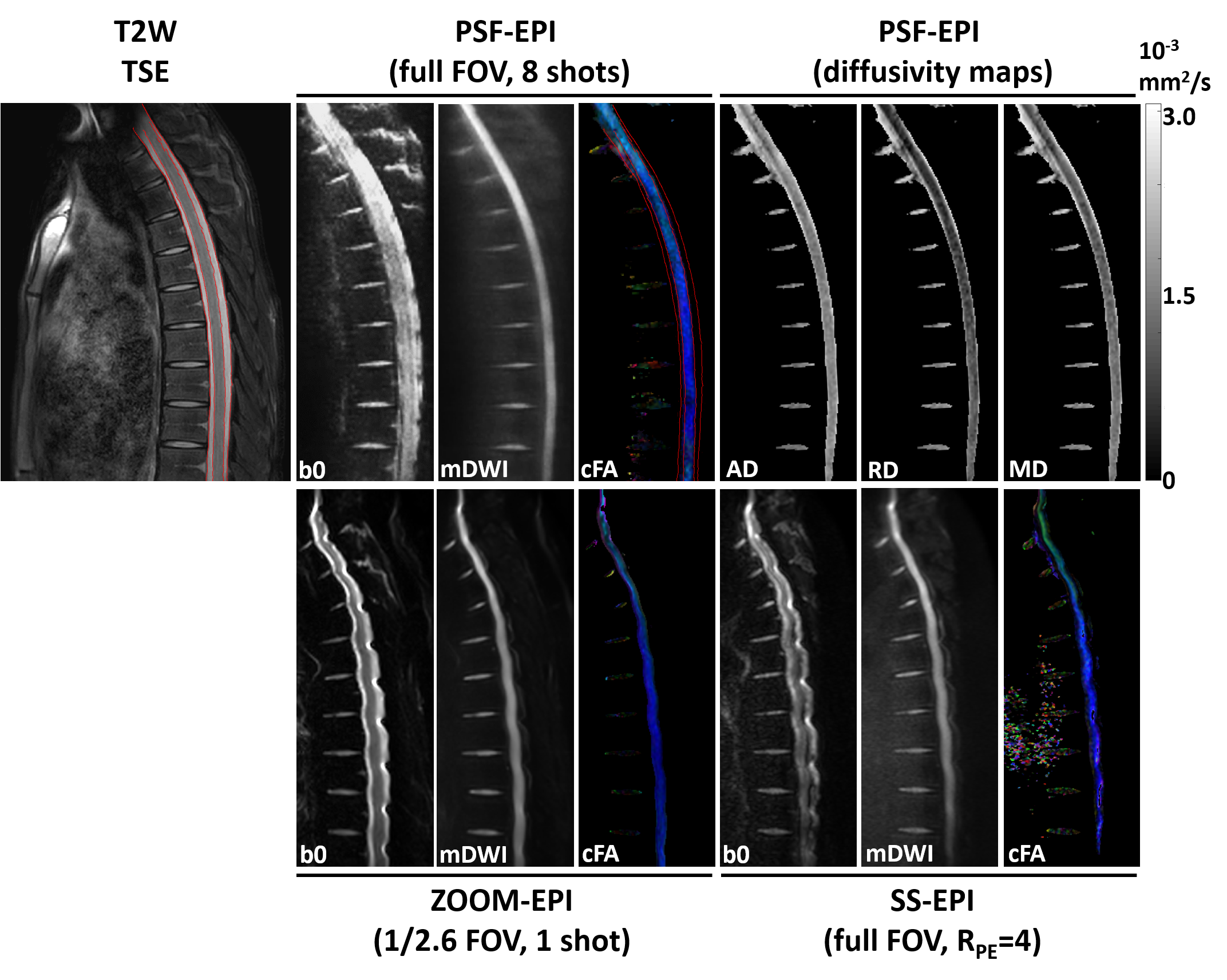
Figure.2 Comparison
between DTI of the thoracic spinal cord using (upper row) full FOV PSF-EPI (8
shots), (lower row) rFOV ZOOM-EPI (1/2.6 FOV, single shot), and full FOV SS-EPI
(RPE=4). Detailed
imaging parameters see Table 1, scan 3-5. Color-encoded FA map (cFA), non-DWI
image (b0) and mean-DWI (mDWI) image are shown. For better comparison, the
major structural boundaries extracted from the T2W reference were overlaid on
the cFA using PSF-EPI. Additionally, the diffusivity maps using PSF-EPI are
also demonstrated.
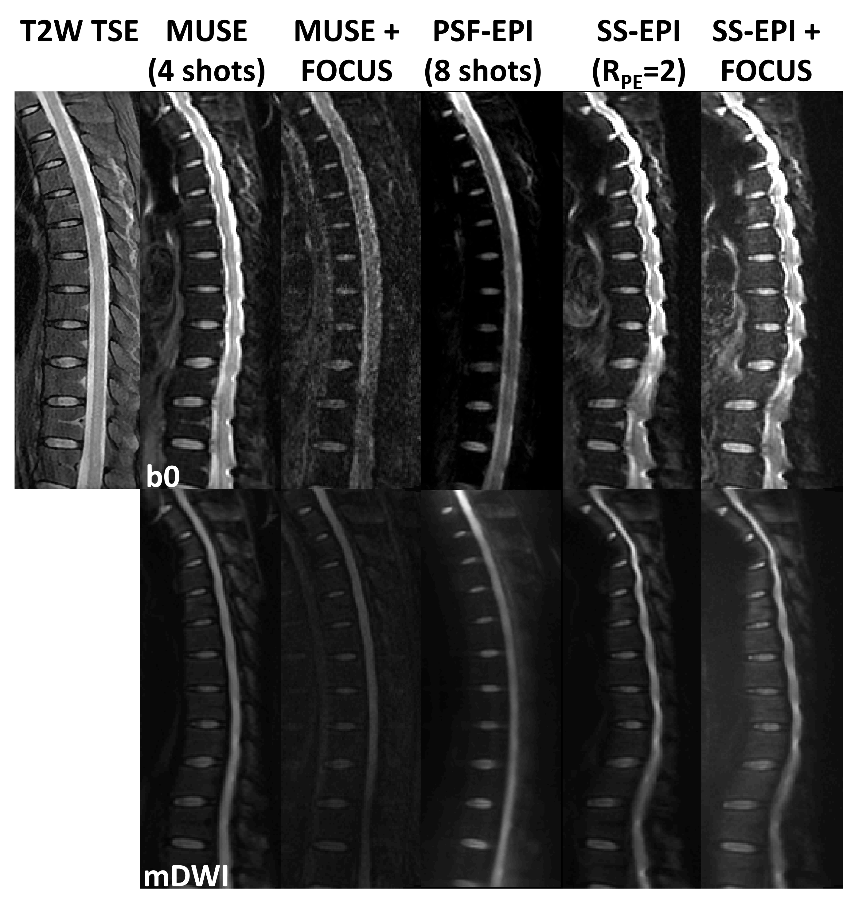
Figure.4 Comparison
of EPI-based methods for distortion correction in thoracic DTI. From left to right:
T2W TSE, 4-shot MUSE with full FOV (Table 1, scan 6), 4-shot MUSE+FOCUS with
1/2 FOV (scan 7), 8-shot PSF-EPI (scan 8), SS-EPI with full FOV and RPE=2 (scan 9),
SS-EPI+FOCUS with 1/2 FOV (scan 10). Upper: T2W TSE reference and non-DWI
images. Lower: mDWI images.