Geraline Vis1, Markus Nilsson1, and Filip Szczepankiewicz1
1Diagnostic Radiology, Clinical Sciences Lund, Lund University, Lund, Sweden
1Diagnostic Radiology, Clinical Sciences Lund, Lund University, Lund, Sweden
Combining
spherical tensor encoding, ultrahigh b-values, and super-resolution
reconstruction enables high-resolution dot fraction imaging in human brain with improved
contrast compared to conventional imaging, where the dot fraction is believed to resemble densely packed cells.
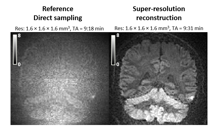
Figure 4 Diffusion weighted imaging with spherical encoding
at b = 4 ms/μm2 in coronal view. A vastly higher contrast is
observed between the cerebellar cortex and white matter using super-resolution
reconstruction (right, contrast ratio 1.82) compared to direct high-resolution sampling
(left, contrast ratio 1.06).
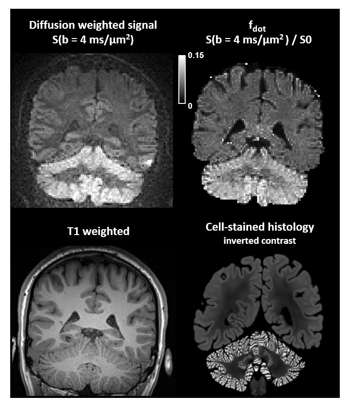
Figure 5 Signal retention using diffusion-weighted imaging
with spherical encoding at b = 4 ms/μm2 (upper left) and estimation of $$$f_{dot}$$$ (upper right) show agreement with cell-stained histology (lower
right plot shows human brain histology from the BigBrain atlas [15]). As
expected, regions of high signal correspond to the cerebellar cortex where
granule cells are densely packed, whereas the white matter is suppressed by the
spherical diffusion encoding (lower left for morphological reference).