Daan Christiaens1,2, Maximilian Pietsch1, Lucilio Cordero-Grande1, Anthony N Price1,3, Jana Hutter1,3, Emer Hughes1, Serena J Counsell1, Joseph V Hajnal1,3, and J-Donald Tournier1,3
1Centre for the Developing Brain, School of Biomedical Engineering and Imaging Sciences, King's College London, London, United Kingdom, 2Department of Electrical Engineering (ESAT-PSI), KU Leuven, Leuven, Belgium, 3Biomedical Engineering Department, School of Biomedical Engineering and Imaging Sciences, King's College London, London, United Kingdom
1Centre for the Developing Brain, School of Biomedical Engineering and Imaging Sciences, King's College London, London, United Kingdom, 2Department of Electrical Engineering (ESAT-PSI), KU Leuven, Leuven, Belgium, 3Biomedical Engineering Department, School of Biomedical Engineering and Imaging Sciences, King's College London, London, United Kingdom
We extended slice-to-volume reconstruction to multi-echo diffusion MRI and applied it in fetal brain imaging. We then explored the combined diffusion-T2* signal characteristics across gestational age.
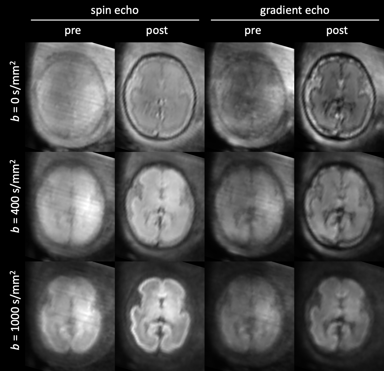
Figure 2: Example of slice-to-volume reconstruction (SVR) in a fetus (28 wPMA) with severe head motion. The images show the mean of each shell before and after SVR, in the spin echo and gradient echo contrast. Grayscale values are min-max normalised per row.
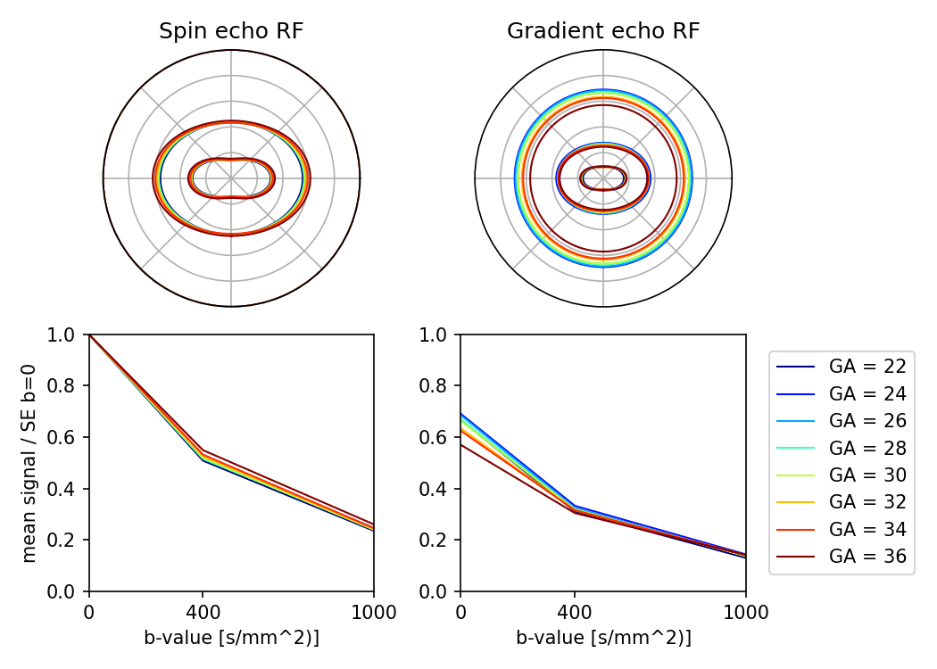
Figure 4: Age trends in the mean tissue response function (RF) in the parenchyma. Tissue RFs for each subject were averaged across bi-weekly age intervals. The spin echo RF shows increasing signal anisotropy and stronger attenuation at young gestational age, as expected. On the other hand, the gradient echo shows stronger T2*-weighting at later gestational ages, most prominently in the b=0 signal.