Beata Bachrata1,2, Bernhard Strasser1,3, Wolfgang Bogner1, Albrecht Ingo Schmid4, Radim Korinek5, Martin Krššák1,2,6, Siegfried Trattnig1,2, and Simon Daniel Robinson1,7,8
1High Field MR Centre, Department of Biomedical Imaging and Image-Guided Therapy, Medical University of Vienna, Vienna, Austria, 2Karl Landsteiner Institute for Clinical Molecular MR in Musculoskeletal Imaging, Vienna, Austria, 3Athinoula A. Martinos Center for Biomedical Imaging, Department of Radiology, Massachusetts General Hospital, Harvard Medical School, Boston, MA, United States, 4High Field MR Centre, Center for Medical Physics and Biomedical Engineering, Medical University of Vienna, Vienna, Austria, 5Institute of Scientific Instruments of the CAS, Brno, Czech Republic, 6Department of Internal Medicine III, Division of Endocrinology and Metabolism, Medical University of Vienna, Vienna, Austria, 7Centre of Advanced Imaging, University of Queensland, St. Lucia, Australia, 8Department of Neurology, Medical University of Graz, Graz, Austria
1High Field MR Centre, Department of Biomedical Imaging and Image-Guided Therapy, Medical University of Vienna, Vienna, Austria, 2Karl Landsteiner Institute for Clinical Molecular MR in Musculoskeletal Imaging, Vienna, Austria, 3Athinoula A. Martinos Center for Biomedical Imaging, Department of Radiology, Massachusetts General Hospital, Harvard Medical School, Boston, MA, United States, 4High Field MR Centre, Center for Medical Physics and Biomedical Engineering, Medical University of Vienna, Vienna, Austria, 5Institute of Scientific Instruments of the CAS, Brno, Czech Republic, 6Department of Internal Medicine III, Division of Endocrinology and Metabolism, Medical University of Vienna, Vienna, Austria, 7Centre of Advanced Imaging, University of Queensland, St. Lucia, Australia, 8Department of Neurology, Medical University of Graz, Graz, Austria
The proposed Simultaneous Multiple Resonance Frequency imaging (SMURF)
approach achieved similar or better fat-water separation than state-of-the-art techniques in the knee, breasts and abdomen and allowed the
elimination of chemical shift effects in fat-water images.
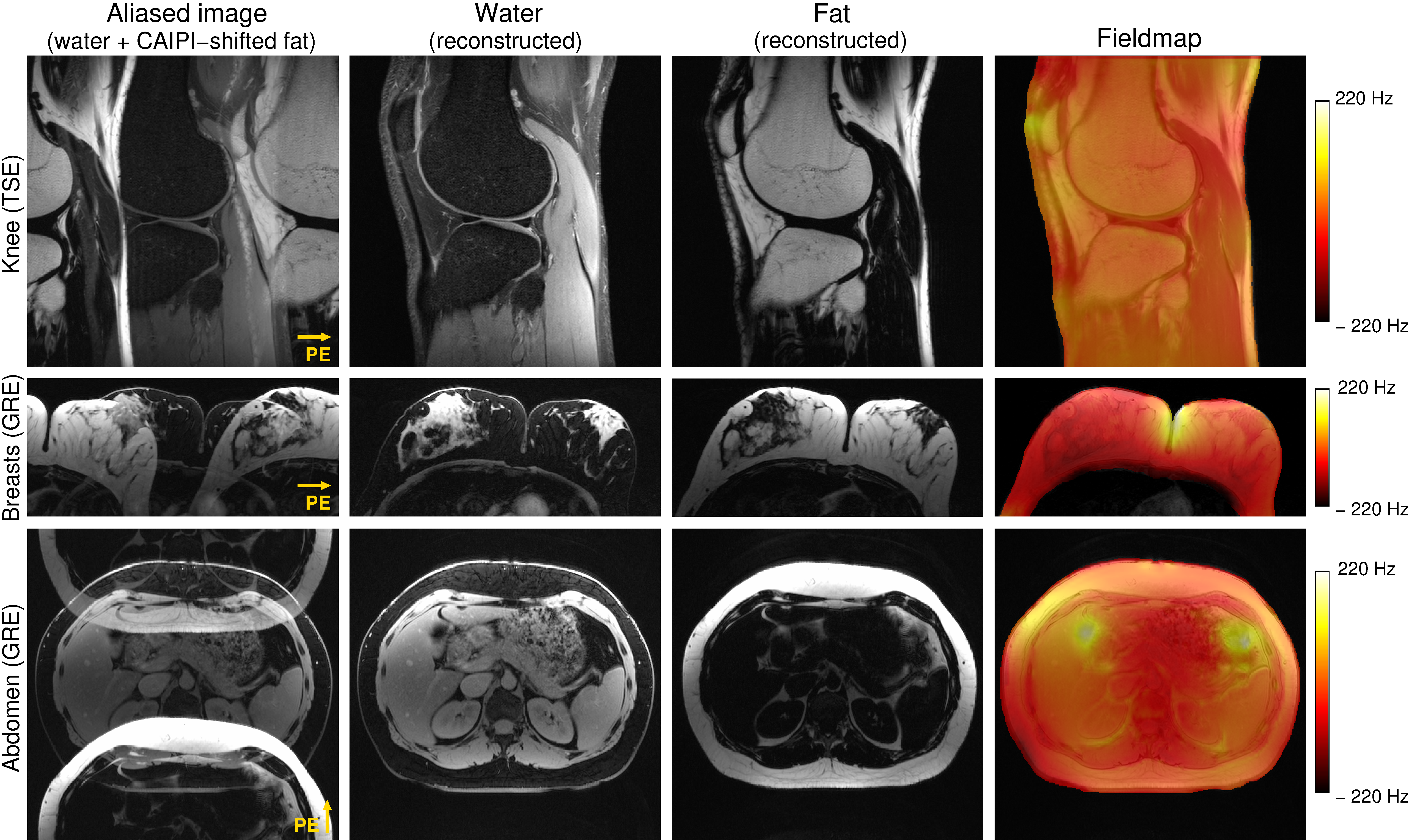
Figure
3: Excitation selectivity and unaliasing quality of SMURF imaging demonstrated
for one exemplary volunteer for each body region under consideration. The
acquired aliased images show the overlapping water and fat images, which are CAIPIRINHA‐shifted
along the phase‐encoding (PE) direction. Separate SMURF water and fat images,
reconstructed from the aliased images, illustrate the achieved separation
quality. (The separated images were rescaled for improved visibility.)
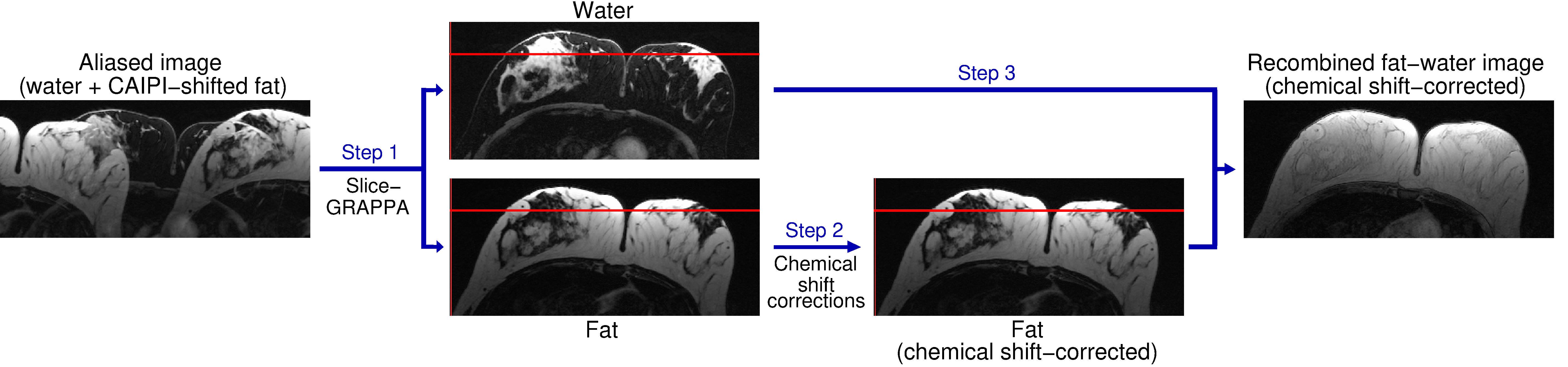
Figure 1: Fat‐water separation, chemical shift corrections, and
recombination in Simultaneous Multiple Resonance Frequency (SMURF) imaging.
Overlapping water and CAIPIRINHA‐shifted fat images are unaliased using
slice‐GRAPPA (Step 1). The fat image is shifted to reverse chemical shift
displacement – see the positions relative to the red reference line – and, for
GRE acquisitions, corrected for chemical shift‐related phase evolution (Step 2).
The fat and water images are then recombined (Step 3), generating a fat‐water
image free of chemical shift artefacts.