Florian Wiesinger1, Timothy Deller2, Floris Jansen2, Jose de Arcos Rodriguez1, Ronny R Buechel3, Philipp A Kaufmann3, and Edwin EGW ter Voert3
1GE Healthcare, Munich, Germany, 2GE Healthcare, Waukesha, WI, United States, 3Department of Nuclear Medicine, University Hospital Zurich, Zurich, Switzerland
1GE Healthcare, Munich, Germany, 2GE Healthcare, Waukesha, WI, United States, 3Department of Nuclear Medicine, University Hospital Zurich, Zurich, Switzerland
The presented method provide an accurate respiratory waveform from listmode PET data. This can then be used for retrospective motion gating of the acquired MR and/or PET data. The method comes for free without requiring an extra motion sensor or complicating the PET/MR imaging workflow.
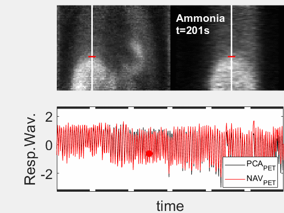
Figure 1 (animated): High temporal-framerate PET
reconstruction (Δt=1s) in the
transient phase ~8mins after the injection of the Ammonia PET tracer.
Corresponding respiratory waveforms (bottom) obtained using either Principal
Component Analysis (PCA, black) or a pencil beam navigator over the lung-liver
interface (red). This patient
demonstrates a deep, regular diaphragmatic breathing pattern.
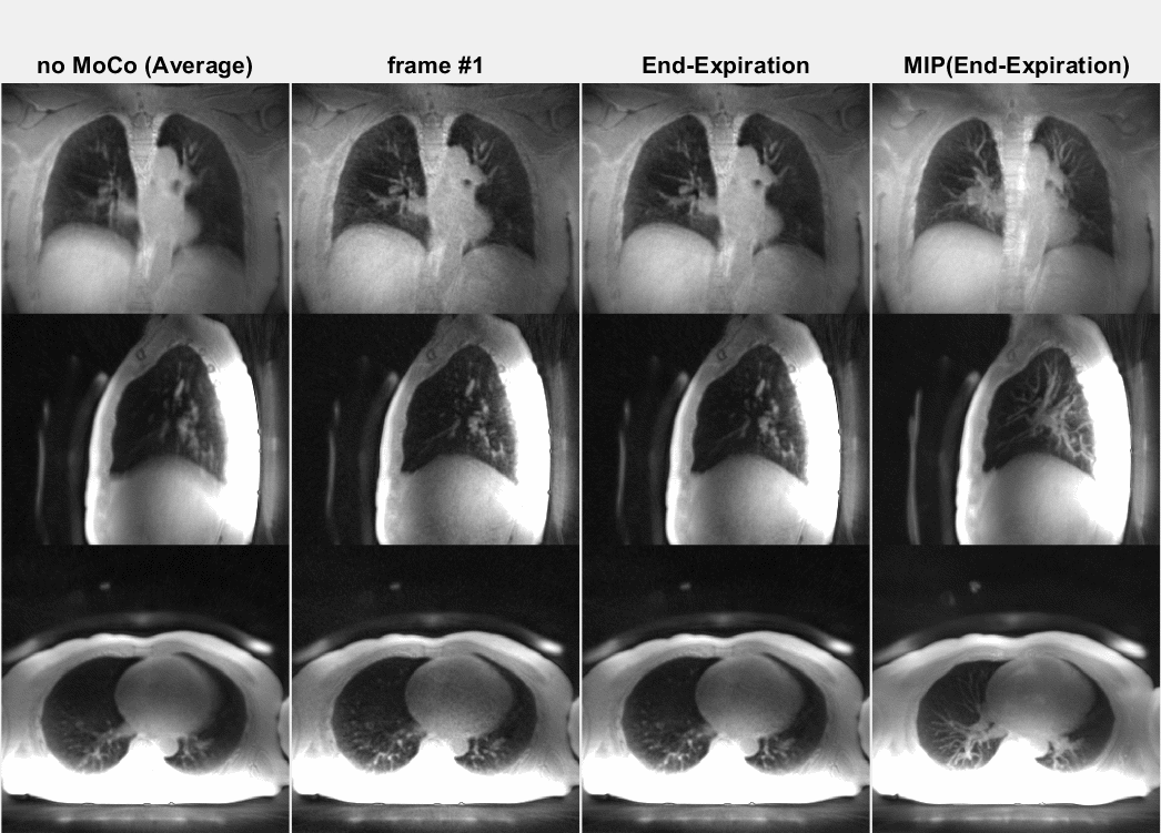
Figure 2 (animated): ZTE lung images corresponding to the Ammonia PET tracer
patient shown in Figure 1. Because of
the deep diaphragmatic breathing the uncorrected (averaged, left) images
show strong motion blurring (especially at the lung-liver interface). Soft-gated
respiratory binning (7 phases, 2nd column) resolves the diaphragmatic
breathing cycle into 7 phases. Most
of the data are acquired in the end-expiratory phase (3rd column) also
providing the sharpest image. Its
Maximum Intensity Project (MIP, right) depicts the vascular anatomy and lung
lesions in fine detail.