Akbar Alipour1, Alan C Seifert1, Bradley Delman1, Raj Shrivastava2, Gregor Adriany3, Zahi Adel Fayad1, and Priti Balchandani1
1Radiology, Icahn School of Medicine at Mount Sinai, New York, NY, United States, 2Neurosurgery, Icahn School of Medicine at Mount Sinai, New York, NY, United States, 3Radiology, University of Minnesota-Medical School, Minneapolis, MN, United States
1Radiology, Icahn School of Medicine at Mount Sinai, New York, NY, United States, 2Neurosurgery, Icahn School of Medicine at Mount Sinai, New York, NY, United States, 3Radiology, University of Minnesota-Medical School, Minneapolis, MN, United States
Results show that the proposed RF resonator sheet can enhance the SNR at the periphery of a RF coil’s region of peak efficiency and sensitivity, thereby extending the anatomical coverage of the commercial MRI head coils at 7T.
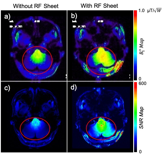
Figure 3: a, b) In-vivo $$$B_1^+$$$ efficiency (normalized by the input power) show a 1.7-fold improvement in the inferior regions of the brain (outlined in red). c, d) In-vivo SNR maps for a subject show that placing the RF sheet enhances the SNR in the cerebellum and brainstem. A 2.0-fold SNR improvement was obtained for the region, outlined in red. The reference voltage was kept constant in with/without modes.
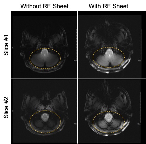
Figure 4: Low flip angle T1- weighted GRE MR images (at two different axial slices) of the lower brain obtained with (right column) and without (left column) the RF sheet show signal enhancement in the inferior regions of the brain improving visibility. Low flip angle sequence was used to avoid RF over-flipping in the close vicinity of the RF sheet.