Yi Chen1, Qi Wang1,2, Hang Zeng1,2, Kengo Takahashi1,2, Sangcheon Choi1,2, Chunqi Qian3, and Xin Yu1,4
1Max Planck Institute for Biological Cybernetics, Tuebingen, Germany, 2Graduate Training Centre of Neuroscience, University of Tuebingen, Tuebingen, Germany, 3Department of Radiology, Michigan State University, East Lansing, MI, United States, 4Athinoula A. Martinos Center for Biomedical Imaging, Massachusetts General Hospital and Harvard Medical School, Charlestown, MA, United States
1Max Planck Institute for Biological Cybernetics, Tuebingen, Germany, 2Graduate Training Centre of Neuroscience, University of Tuebingen, Tuebingen, Germany, 3Department of Radiology, Michigan State University, East Lansing, MI, United States, 4Athinoula A. Martinos Center for Biomedical Imaging, Massachusetts General Hospital and Harvard Medical School, Charlestown, MA, United States
Here, we demonstrate an inductive coil embedded beneath the surface coil
to obtain laminar-specific information for high spatiotemporal resolution (100μm
and 100ms) in both focal rat cortex and multiregional mapping of the brain
functional connectivity with the optogenetic tool.
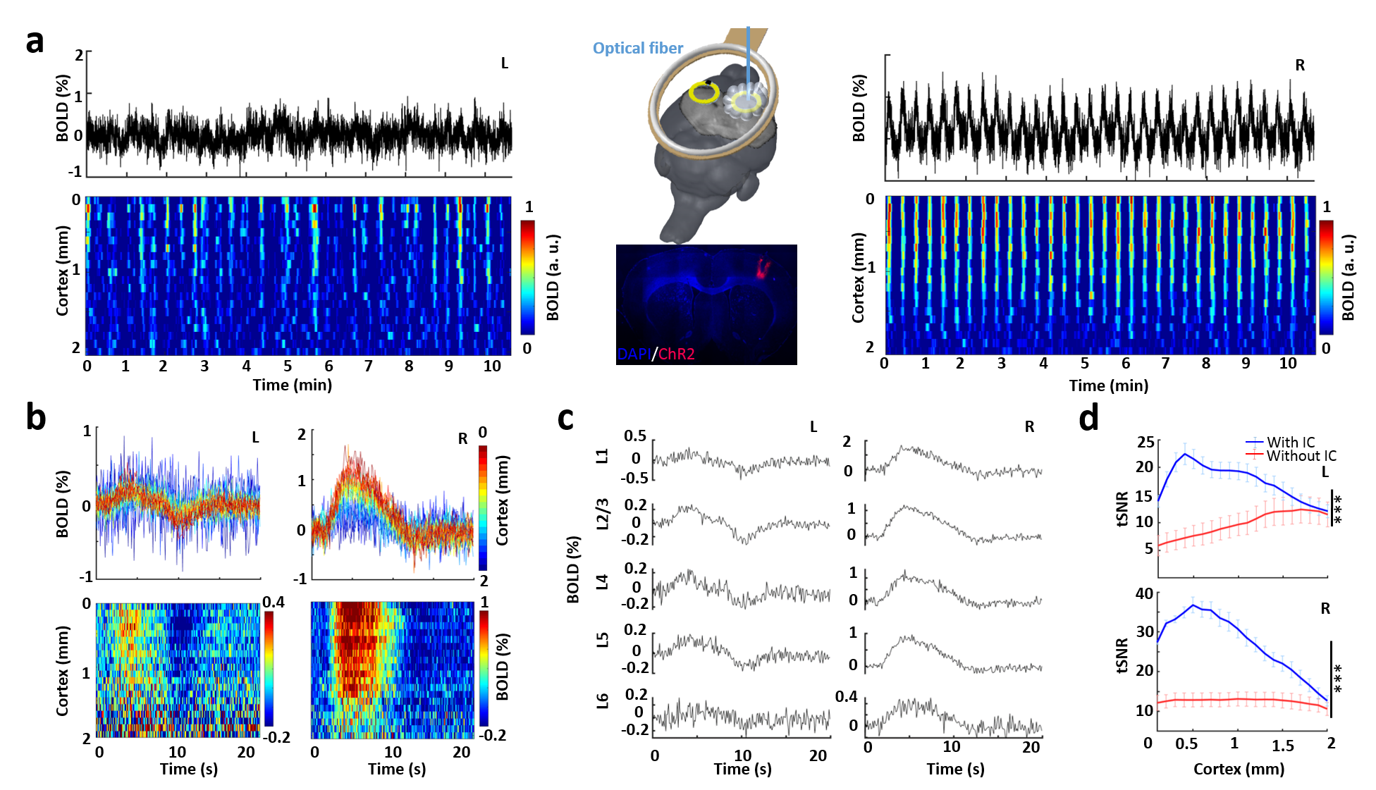
Fig. 3.
Optogenetically evoked BOLD responses in bilateral enhanced FP-S1 regions. a) Time
course and spatiotemporal map of bilateral BOLD responses induced directly from
optogenetic stimulation in the right FP-S1 (right) and projected left FP-S1 (left,
n = 3 rats, 49 trials). b) BOLD-change in each voxel along cortical depth on
both hemispheres (n = 3 rats, 49 trials). c) Different laminar-specific
responses of both hemispheres. d) Significantly higher tSNR with (blue) than
without (red) implanted inductive coils (paired-sample t-test, ***P <0.001,
n = 3 rats, mean ± SEM).
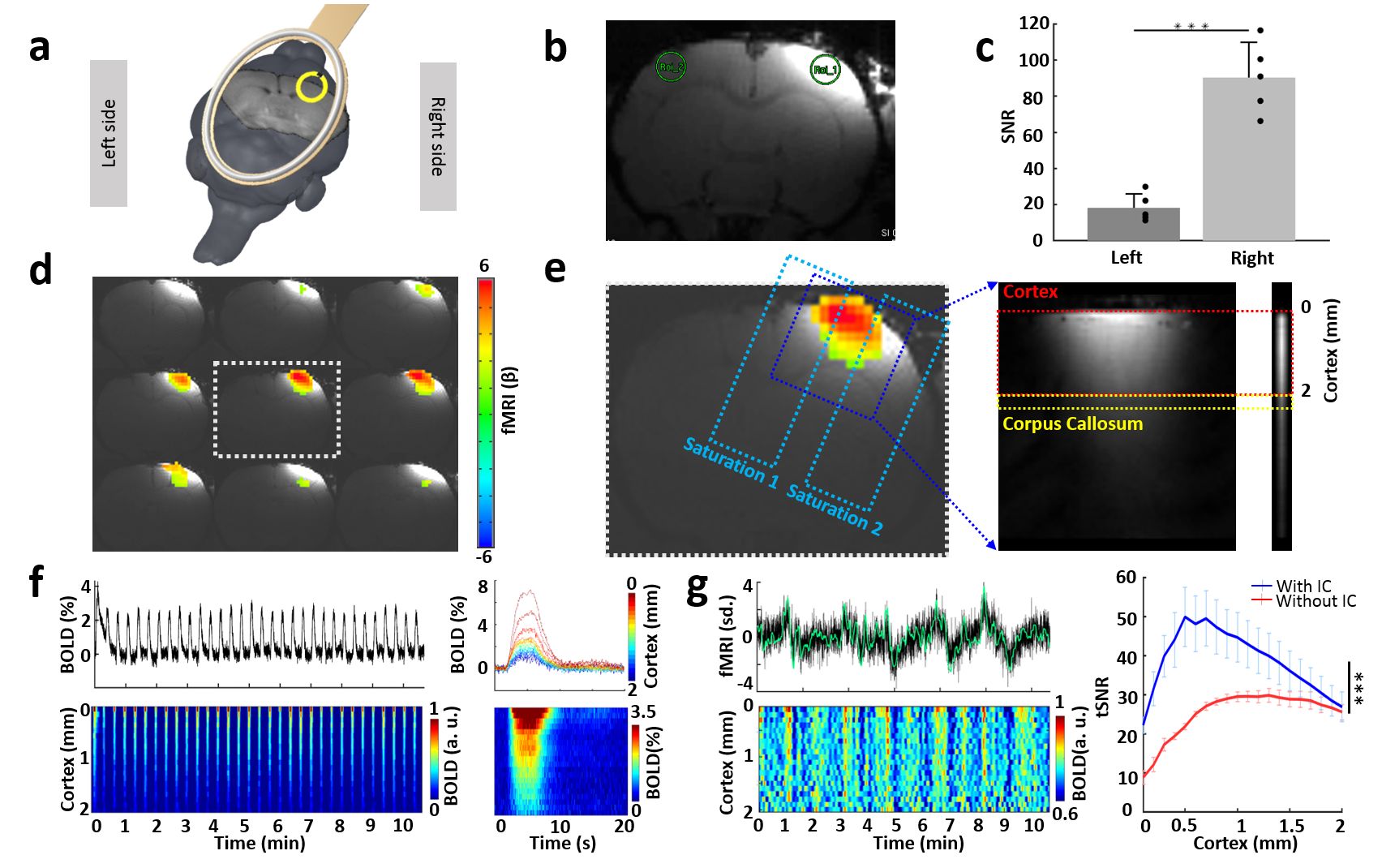
Fig. 2.
BOLD responses detected using a line-scanning method in an enhanced region. a) Experimental
setup. b) Enhanced focal intensity in the right FP-S1. c) Significantly higher
SNR in ROI 1 with the inductive coil. d) BOLD activation map. e) The procedure for line-scanning
method. f) BOLD change and spatiotemporal map in the cortex along the cortical
depth (20 voxels, 2 mm, n = 3 rats). g) Resting-state hemodynamic fluctuation
from right cortex and spatiotemporal map along the cortex. Right, significantly
higher tSNR with the inductive coil (blue) than previous results (red).