Aurelien Destruel1, Ewald Weber1, Mingyan Li1, Jin Jin1,2, Craig Engstrom3, Feng Liu1, and Stuart Crozier1
1School of Information Technology and Electrical Engineering, The University of Queensland, Brisbane, Australia, 2Siemens Healthcare Pty Ltd, Brisbane, Australia, 3School of Human Movement and Nutrition Sciences, The University of Queensland, Brisbane, Australia
1School of Information Technology and Electrical Engineering, The University of Queensland, Brisbane, Australia, 2Siemens Healthcare Pty Ltd, Brisbane, Australia, 3School of Human Movement and Nutrition Sciences, The University of Queensland, Brisbane, Australia
A novel radiofrequency coil element is
presented in numerical simulations (compared with fractionated dipoles) and in
vivo 7T MRI. The proposed design has excellent stability/performance with
fixed matching/tuning across different body regions and volunteers.
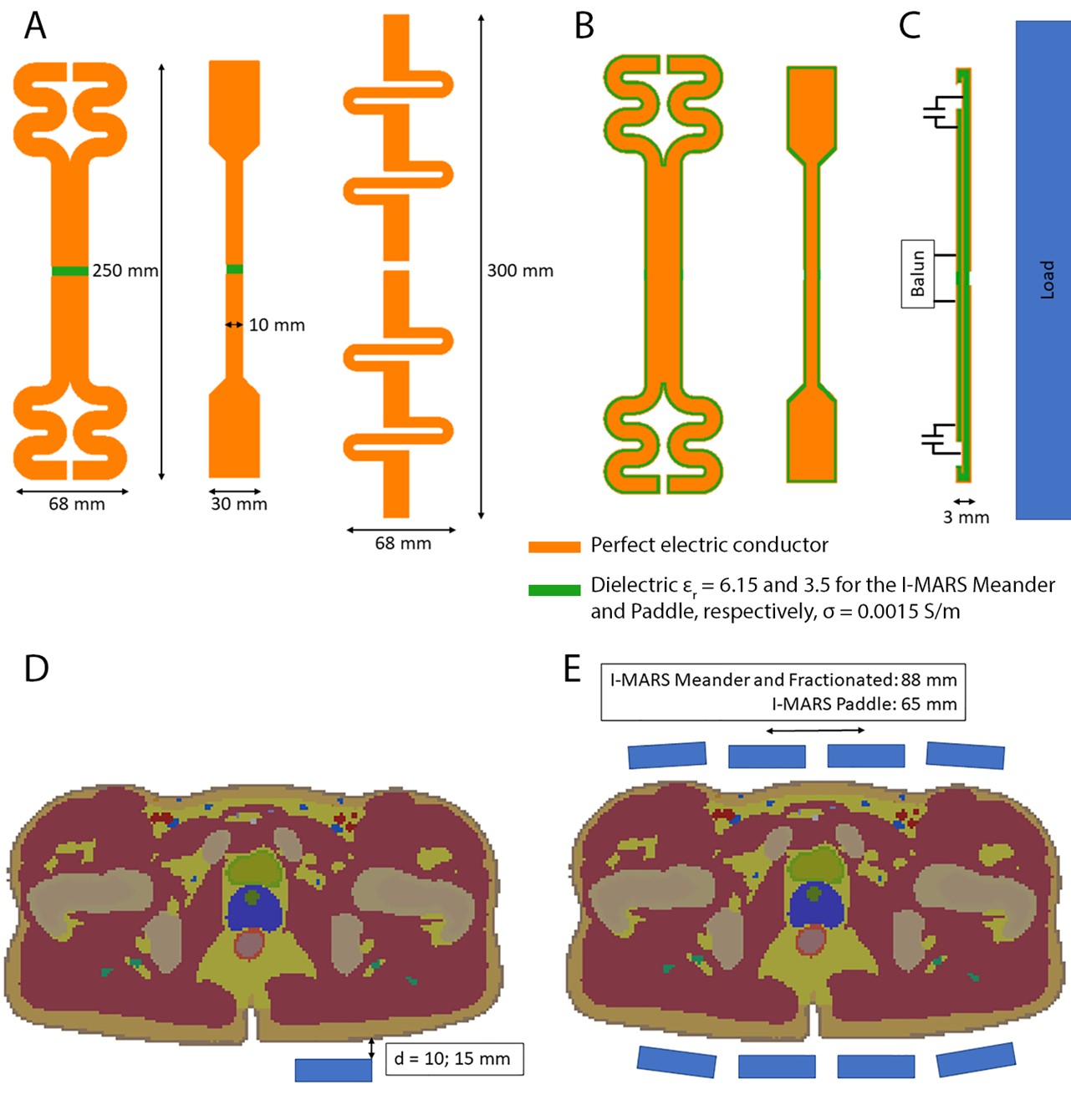
Figure 1: A) Models of the (left) I-MARS
Meander, (center) I-MARS Paddle, (right) Fractionated dipole. The I-MARS Paddle
had a narrower center compared to the ends to achieve a more focused field
distribution.; B) Coronal cut-away views for I-MARS Meander and I-MARS Paddle
models; C) Sagittal cut-away view for the I-MARS Paddle, showing the location
of the tuning capacitors and balun; D) Axial slice of Duke and a coil element
to compare the stabilities of the different coils; E) Axial slice of Duke and
eight coil arrays to compare RF shimming performance, B1 and SAR
efficiency.
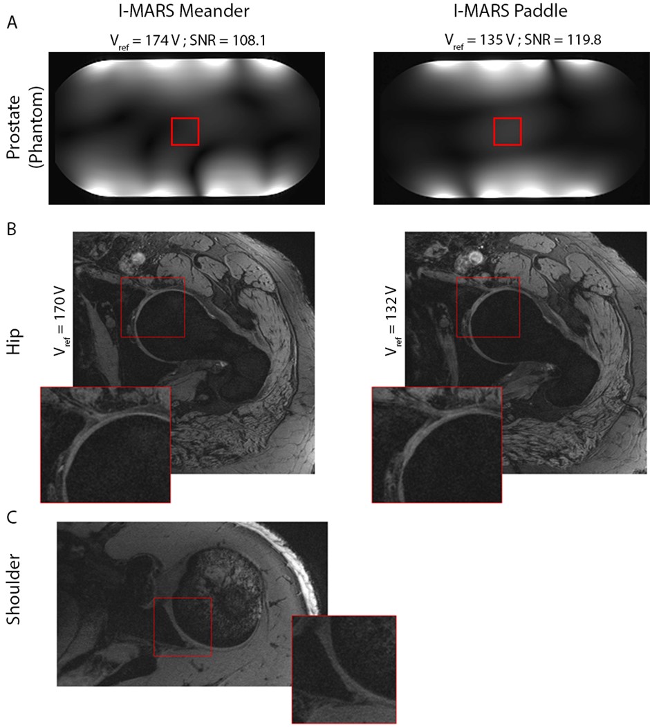
Figure 3: A)
Axial GRE slices of a phantom, with the I-MARS coil arrays in prostate
configuration. The reference voltage (Vref) required to calibrate
the B1 in the ROI (red square) was compared, as well as the SNR in
the ROI. B) we-DESS images of the hip acquired with the I-MARS Meander and
I-MARS Paddle arrays, comparing the Vref required to calibrate the B1
over the femoral head. C) we-DESS images of the shoulder acquired with the
I-MARS Meander array.