Yujin Jung1, Jaeseok Park2, Seong-Gi Kim2, and Sung-Hong Park1
1Department of Bio and Brain engineering, Korea Advanced Institute of Science and Technology, Daejeon, Korea, Republic of, 2Department of Global Biomedical Engineering, Sungkyunkwan University, Suwon, Korea, Republic of
1Department of Bio and Brain engineering, Korea Advanced Institute of Science and Technology, Daejeon, Korea, Republic of, 2Department of Global Biomedical Engineering, Sungkyunkwan University, Suwon, Korea, Republic of
In this study, we developed a new diffusion-weighted steady state CEST sequence using 3D EPI at 7T. The technique was tested in phantom and human brain, and the preliminary CEST-weighted apparent diffusion coefficient maps provided both CEST and diffusion information.
Figure
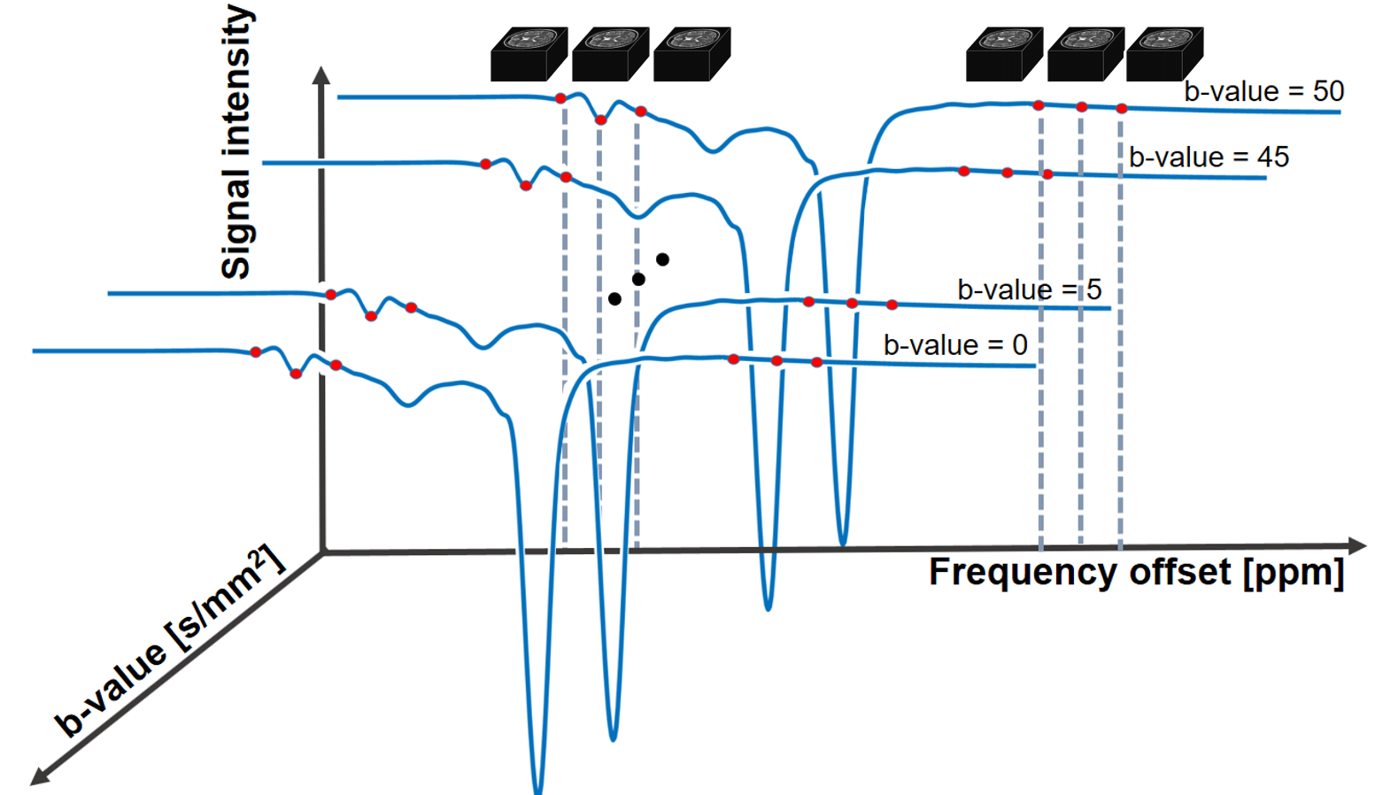
1. Schematic illustration of diffusion-weighted CEST. 3D images were acquired at 6
offsets around 3.5ppm and -3.5ppm (6 point acquisition1) and at 11
b-values = 0, 5, … 45, 50 s/mm2.
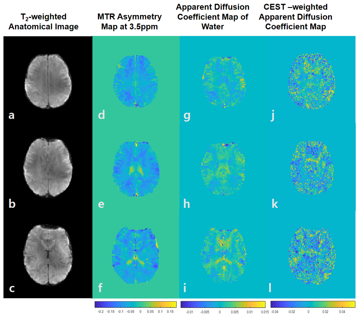
Figure 5.
Diffusion maps of human brain. Axial
anatomical images in three slices (each row) are shown (a, b, c). MTR asymmetry
maps at 3.5 ppm demonstrates CEST distribution in human brain (d, e, f). Apparent
diffusion coefficient map was calculated and the average value was 0.001mm2/s
across the slices (g, h, i). White matter tracks were discernable in the
CEST-weighted apparent diffusion coefficient map (j, k, l).