Daniel J. West1, Lucilio Cordero-Grande1,2,3, Rui P. A. G. Teixeira1,2, Giulio Ferrazzi4, Joseph V. Hajnal1,2, and Shaihan J. Malik1,2
1Biomedical Engineering and Imaging Sciences, King's College London, London, United Kingdom, 2Centre for the Developing Brain, King's College London, London, United Kingdom, 3Biomedical Image Technologies, ETSI Telecomunicación, Universidad Politécnica de Madrid & CIBER-BNN, Madrid, Spain, 4IRCCS San Camilo Hospital, Venice, Italy
1Biomedical Engineering and Imaging Sciences, King's College London, London, United Kingdom, 2Centre for the Developing Brain, King's College London, London, United Kingdom, 3Biomedical Image Technologies, ETSI Telecomunicación, Universidad Politécnica de Madrid & CIBER-BNN, Madrid, Spain, 4IRCCS San Camilo Hospital, Venice, Italy
DISORDER k-space
trajectories can be used for motion corrected MR fingerprinting, here applied
to ihMT measurement. Resultant parameter maps allow quantification of dipolar
relaxation times in vivo and support future use of this motion
correction framework for general MRF-style methods.
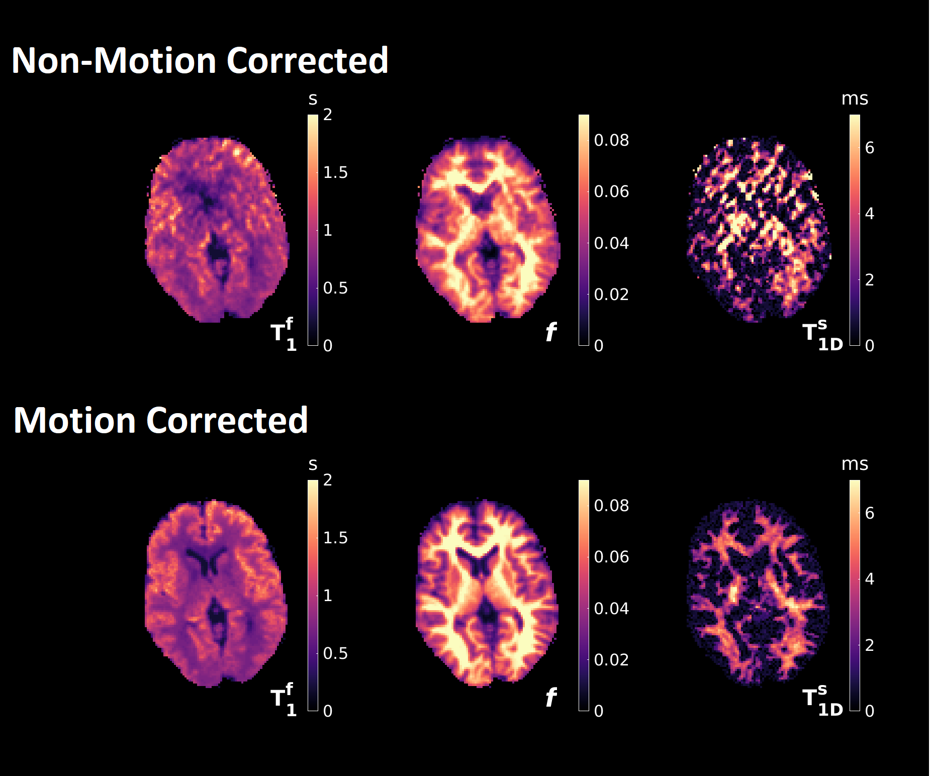
Figure 5:
Dictionary fits to in vivo data with and without motion correction. Motion artefacts are significantly reduced for
the former, enabling quantification of T1Ds. The lower contrast-to-noise
ratio of ihMT compared to MT means quantification of dipolar parameters is hindered prior to
correction. GM-WM contrast appears enhanced
for T1Ds versus f and highly myelinated structures
become more discernible. Semisolid relaxation times were fixed at T1Zs = 0.2s and T2s = 7.5μs; free pool T2 at T2f = 69ms; exchange rate at K = 50s-1 and main magnetic field induced phase at
ΔB0 = 013,14.
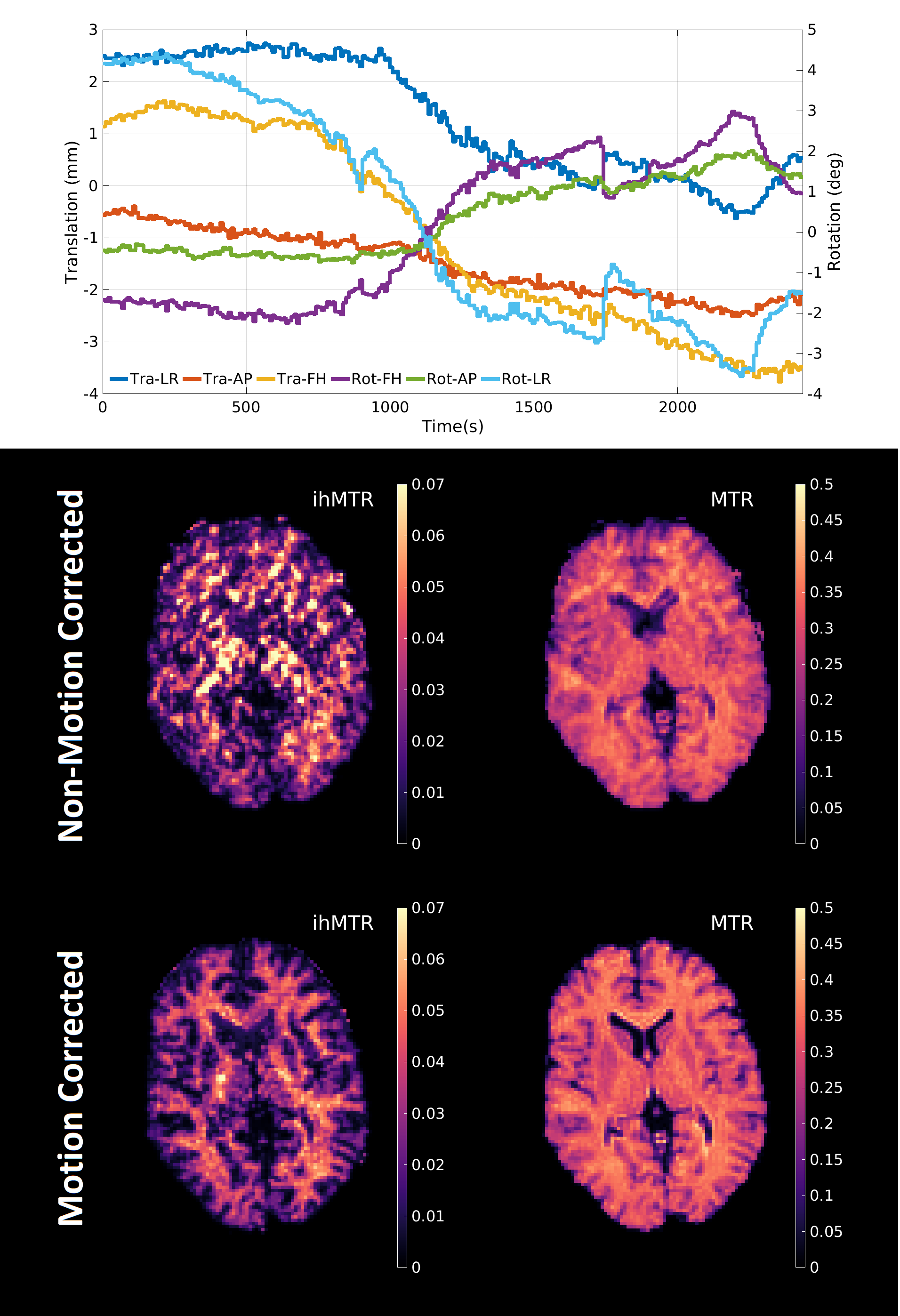
Figure 4: Top: Estimated motion traces during the in
vivo acquisition. Results are from a compliant volunteer who was instructed not to move during the acquisition. "Tra" refers to
transversal motion and "Rot" refers to rotation; "LR"
indicates the left-right direction, "AP" is anterior-posterior, and "FH"
is foot-head. Bottom: Example in
vivo contrast ratio maps with and without motion correction from a central
axial slice and reconstructed using the first eight singular components. MTR
shows strong grey matter (GM)-white matter (WM) contrast and ihMTR is more
correlated to WM.