Muriah D Wheelock1, Patricia Mansfield2, Jeremy F Strain1, Beau M Ances1, Oliver Preische3, John C Morris1, Randall J Bateman1, Mathias Jucker3, Tammie L.S. Benzinger1, Adam T Eggebrecht1, and Brian A Gordon1
1Washington University in St. Louis, St. Louis, MO, United States, 2St. Louis University, St. Louis, MO, United States, 3University of Tubingen, Tubingen, Germany
1Washington University in St. Louis, St. Louis, MO, United States, 2St. Louis University, St. Louis, MO, United States, 3University of Tubingen, Tubingen, Germany
For the first time we demonstrate that blood serum Neurofilament light (NfL) is associated with concurrent functional connectivity (FC) of the default mode network (DMN) and FC between the DMN and control networks. NfL, amyloid, and DMN FC are predictive of concurrent cognitive function.
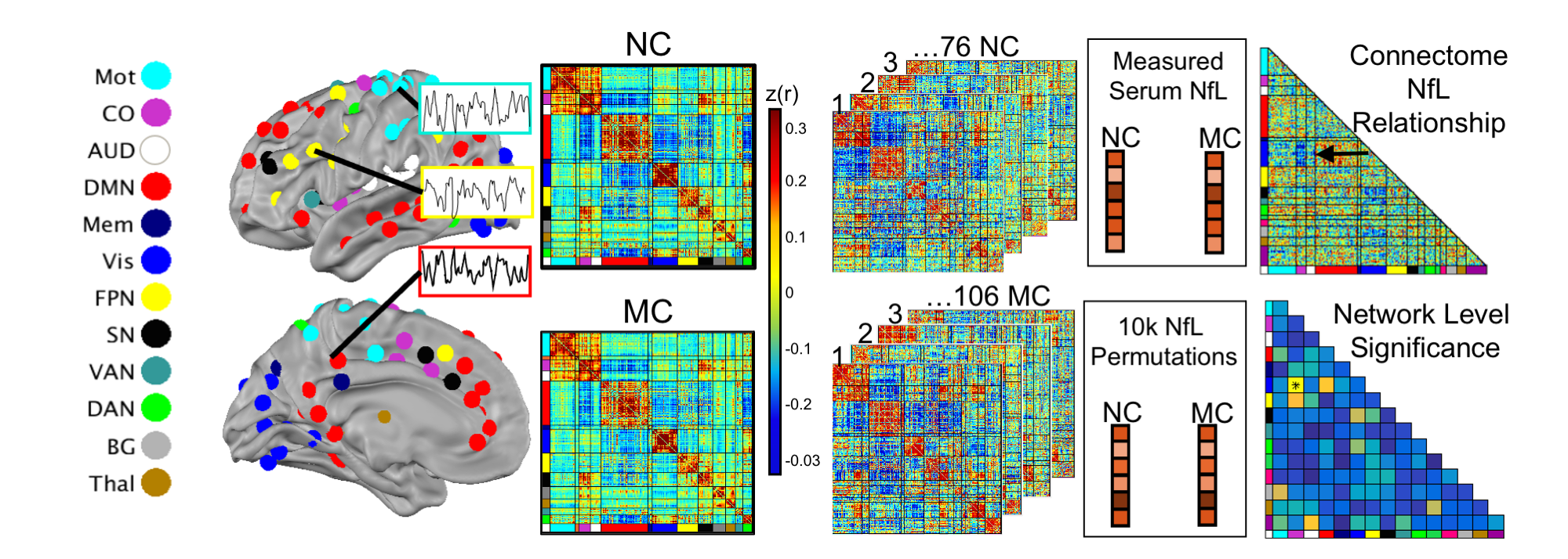
Figure 1. Network Level Analysis. After preprocessing and motion correction, BOLD time series were extracted from 246 spherical regions of interest. Nodes [MW1] of the same color belong to the same brain network community. The non-parametric correlation between NfL and whole brain connectome was examined separately for each group. Significance was established by randomly permuting the NfL values 10,000 times and measuring the permuted connectome-NfL relationship. [MW1]Adam wants me to include the actual data from this study in the cartoon
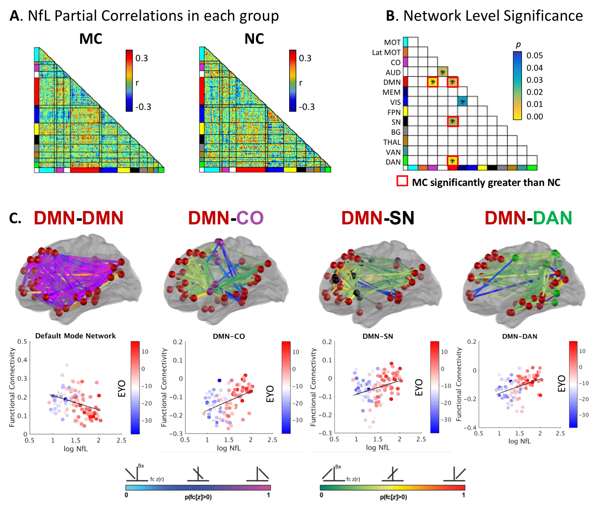
Figure 3. Networks associated with NfL. A) Partial correlations between NfL and FC in MC and NC groups. B) MC demonstrated stronger associations with NfL than NC in four network pairs (red boxes, p<0.05). C) The pink to light blue spectrum indicates a negative correlation between NfL and FC wherein individuals with the highest NfL have the lowest DMN FC (pink). The red to green spectrum indicates a positive correlation between NfL and FC wherein individuals with the highest (green) NfL have reduced anti-correlation between DMN and SN, DAN, and CO networks.
