Lingxiao Cao1, Hailong Li1, Jing Liu1, Xue Li2, Suming Zhang1, Xinyu Hu1, Qiyong Gong1, and Xiaoqi Huang1
1Huaxi MR Research Center (HMRRC), Functional and molecular imaging Key Laboratory of Sichuan Province, Department of Radiology, West China Hospital, Sichuan University, Chengdu, China, 2Sichuan University, Chengdu, China
1Huaxi MR Research Center (HMRRC), Functional and molecular imaging Key Laboratory of Sichuan Province, Department of Radiology, West China Hospital, Sichuan University, Chengdu, China, 2Sichuan University, Chengdu, China
Connectivity-based parcellation
revealed distinct functional connectivity profiles of thalamic subdivisions
between the OCD patients and HC.
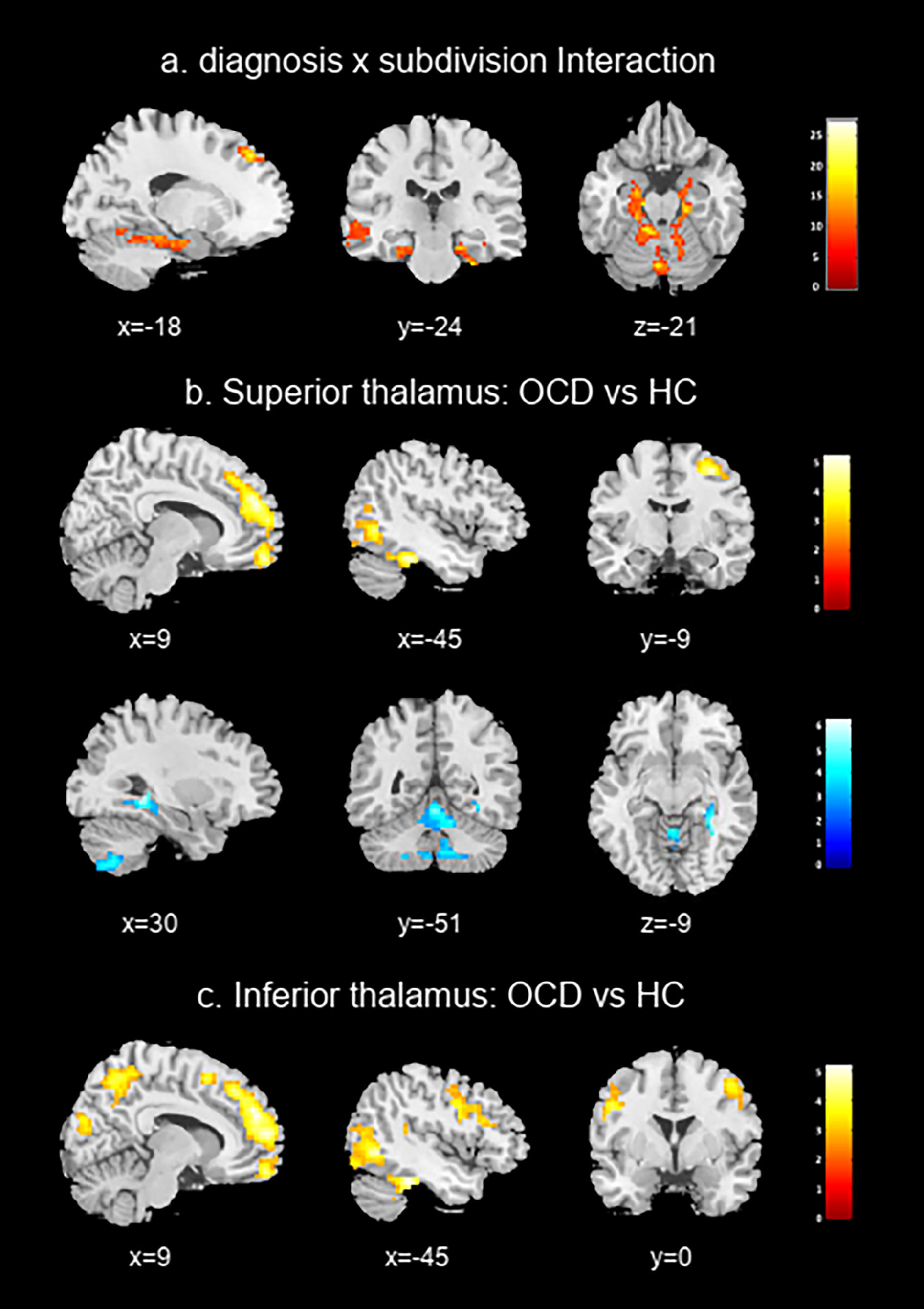
Figure 3 Brain regions
exhibiting significant interaction (a), and increased functional connectivity
in OCD compared with HC (shown in warm color) and HC compared with OCD (shown
in cool color) with superior thalamus (b), and inferior thalamus (c).
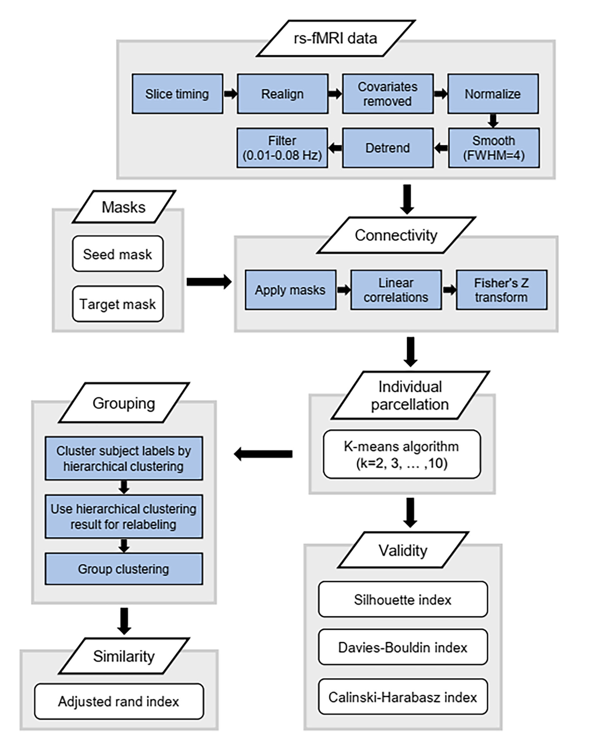
Figure 1 A flowchart of resting-state
functional MRI (rs-fMRI) data analysis and thalamic parcellation.