Xiaole Zhong1 and J. Jean Chen1,2
1Rotman Research Institute, Baycrest Health Sciences, Toronto, ON, Canada, 2Department of Medical Biophysics, University of Toronto, Toronto, ON, Canada
1Rotman Research Institute, Baycrest Health Sciences, Toronto, ON, Canada, 2Department of Medical Biophysics, University of Toronto, Toronto, ON, Canada
The frequency of the resting-state fMRI signal increases with age while standard deviation decreases. This behaviour is more pronounced in 0.1-0.3Hz than in 0.01-0.1 Hz.
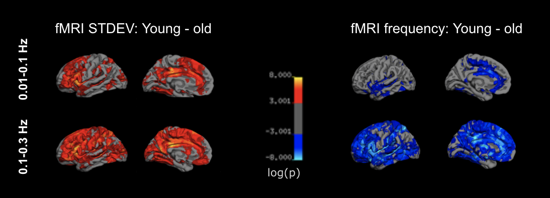
Figure 2. rs-fMRI frequency versus age. Only significant regions are shown in colour. fMRI fluctuation frequency is higher in the older adults, also with the 0.1-0.3 Hz band showing more widespread differences. All cases show significant differences in the cingulate cortex and paracentral cortex.
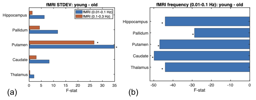
Figure 4. rs-fMRI amplitude and frequency versus age: subcortical regions. (a) The fMRI signal amplitude shows significant age effects (young > old) in the putamen, in both fMRI frequency bands. (b) The fMRI frequency is higher in the older adults in all subcortical structures, but only in the 0.01-0.1 Hz band. Significance is indicated by asterisks.