Christopher R. Kouyoumdjian1, Kayley Marchena2,3, Masud Hussain3, and Bradley J. MacIntosh2,3
1University of Toronto, Scarborough, ON, Canada, 2Department of Medical Biophysics, University of Toronto, Toronto, ON, Canada, 3Sunnybrook Research Institute, Toronto, ON, Canada
1University of Toronto, Scarborough, ON, Canada, 2Department of Medical Biophysics, University of Toronto, Toronto, ON, Canada, 3Sunnybrook Research Institute, Toronto, ON, Canada
BMI is an impactful predictor of bilateral hippocampal T2* estimates in diabetes-related cohorts. BMI was associated with resting state fALFF activation in the caudate nuclei and thalamic nuclei.
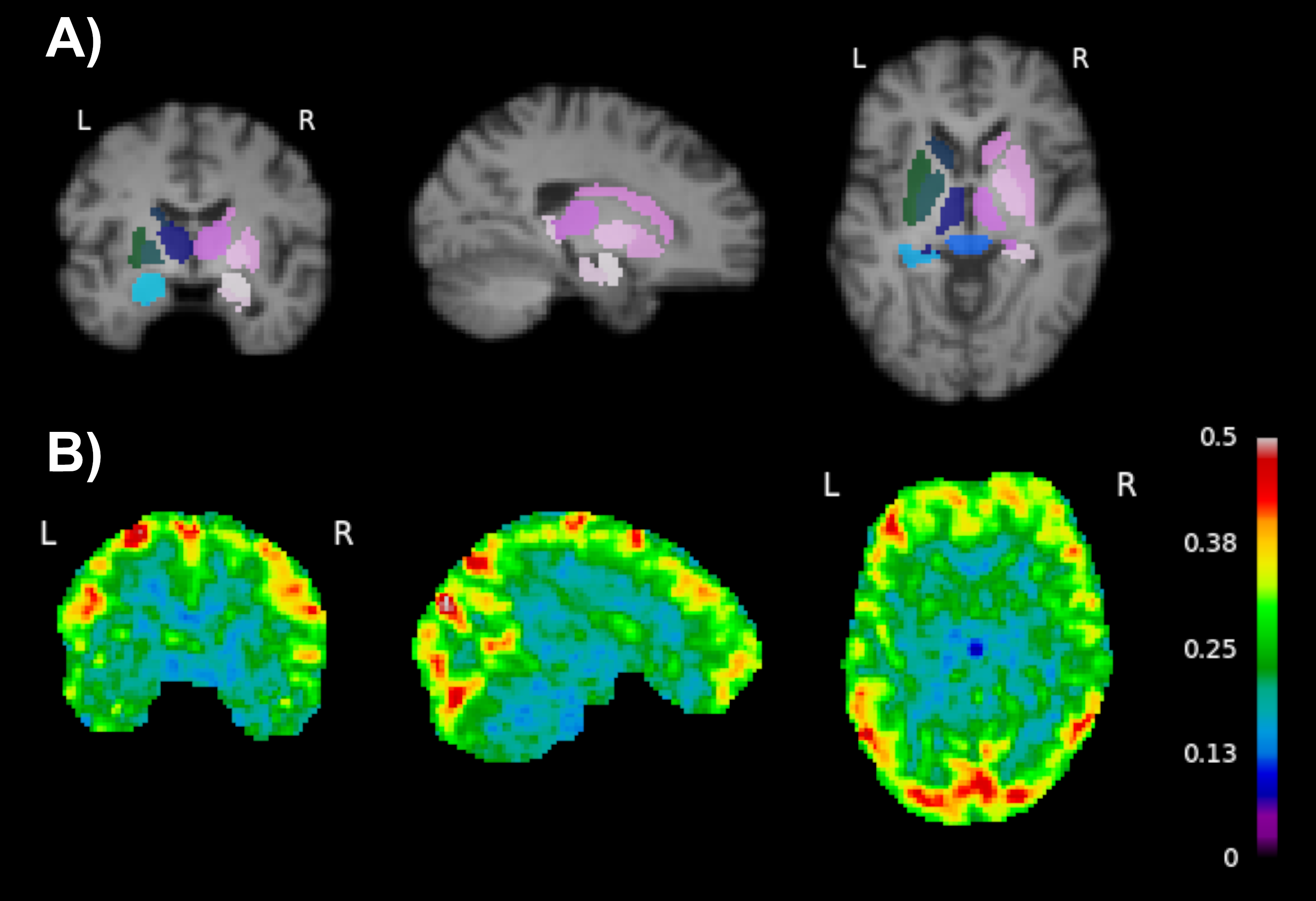
Figure 2: Visualization of subcortical masks (A) and whole-brain fALFF maps (B). Coronal, sagittal, and axial views are in MNI coordinate space (x=16, y=15, z=6). The colours in (B) show the fraction of the total power low frequency fluctuations within the 0.01–0.1 Hz frequency bands.
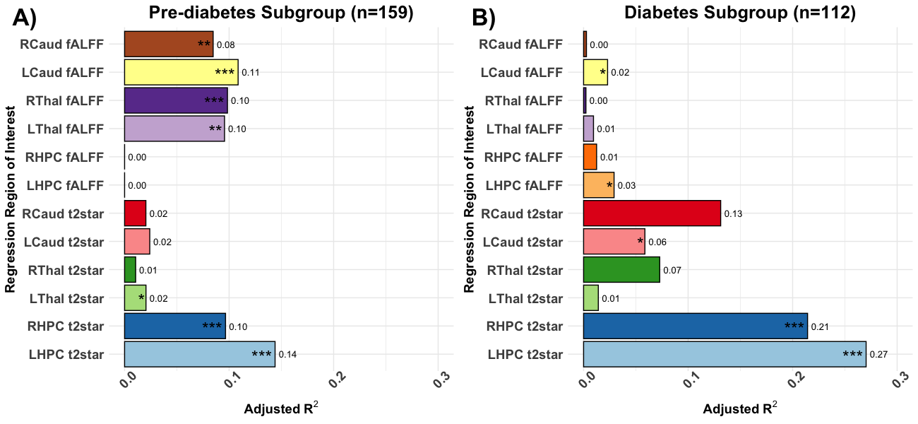
Figure 3: A summary of the multiple regression models for each region of interest across the pre-diabetes (A) and diabetes (B) cohorts. The model included age, sex, and BMI as covariates. Models where BMI significantly predicted the outcome variable were denoted as follows: * P<.05; ** P<.01; *** P<.001. Caud: caudate nucleus, HPC: hippocampus, Thal: thalamus, L/R: left/right hemisphere.