Russell W. Chan1,2, Ji Won Bang2, Vivek Trivedi2, Peiying Liu3, Gadi Wollstein2, Joel S. Schuman2, and Kevin C. Chan1,2,4
1Neuroscience Institue, New York University Grossman School of Medicine, New York, NY, United States, 2Department of Ophthalmology, New York University Grossman School of Medicine, New York, NY, United States, 3Department of Radiology, Johns Hopkins University School of Medicine, Baltimore, MD, United States, 4Department of Radiology, New York University Grossman School of Medicine, New York, NY, United States
1Neuroscience Institue, New York University Grossman School of Medicine, New York, NY, United States, 2Department of Ophthalmology, New York University Grossman School of Medicine, New York, NY, United States, 3Department of Radiology, Johns Hopkins University School of Medicine, Baltimore, MD, United States, 4Department of Radiology, New York University Grossman School of Medicine, New York, NY, United States
Whole-brain cerebrovascular
reactivity (CVR) assessments in
glaucoma patients have been lacking. We applied relative
CVR (rCVR) mapping using resting-state fMRI. We found visual cortical rCVR decreases
with glaucoma severity and is coupled with clinical ophthalmic assessments.
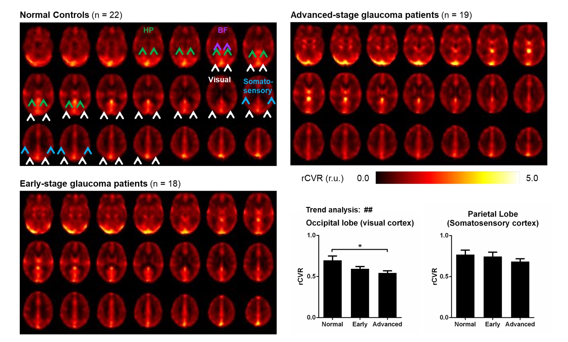
Figure 2: Advanced-stage glaucoma patients have
significantly lower rCVR in visual cortex. Averaged rCVR maps were calculated for normal controls, early-stage glaucoma patients,
and advanced-stage glaucoma patients. Advanced-stage glaucoma patients have
significantly lower rCVR in the visual cortex. In addition, rCVR in the visual
cortex has a decreasing trend with glaucoma severity. These were not observed
in the somatosensory cortex. One-way ANOVA followed by Bonferroni’s post hoc
test (*p<0.05) and trend analysis (##p<0.01) were applied. Error bars indicate ±SEM.
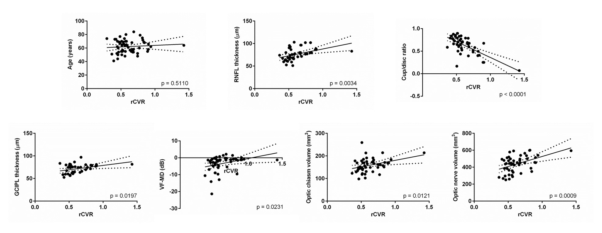
Figure 3: Visual cortical rCVR is coupled with clinical
ophthalmic assessments and volumetric MRI assessments. Scatter plots show the
relationship between rCVR in visual cortex and age, clinical ophthalmic
assessments, as well as volumetric MRI assessments. Visual cortical rCVR is
positively correlated with RNFL thickness, GCIPL thickness, VF-MD, optic chiasm
volume and optic nerve volume, while negatively correlated with cup/disc ratio.
No significant correlation was found between visual cortical rCVR and age.
Linear regression was applied.