Siqi Cai1,2, Zhifeng Shi3, Yuchao Liang4, Chunxiang Jiang1,2, Shihui Zhou1,2, and Lijuan Zhang*1
1Shenzhen Institutes of Advanced Technology, Chinese Academy of Sciences, Shenzhen, China, 2University of Chinese Academy of Sciences, Beijing, China, 3Huashan Hospital of Fudan University, Shanghai, China, 4Neurosurgery, Beijing Tiantan Hospital of Capital Medical University, Beijing, China
1Shenzhen Institutes of Advanced Technology, Chinese Academy of Sciences, Shenzhen, China, 2University of Chinese Academy of Sciences, Beijing, China, 3Huashan Hospital of Fudan University, Shanghai, China, 4Neurosurgery, Beijing Tiantan Hospital of Capital Medical University, Beijing, China
Cerebral gliomas induced alteration in dFC featuring more frequent strengthened connectivity and state transition between strong and sparse functional connectivity. This provides a new biomarker for the tumor characterization of glioma.
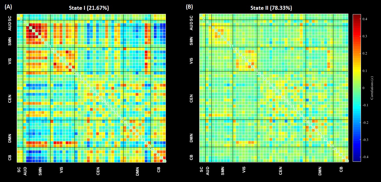
Figure 2. The cluster centroids of two functional connectivity states. (SC: subcortical, AUD: auditory network, SMN: sensorimotor network, VIS: visual network, CEN: cognitive executive network, DMN: default mode network, CB: cerebellum)
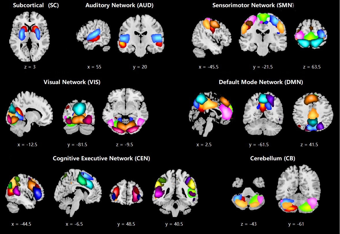
Figure 1. Spatial maps of 49 selected independent components (IC). Colors discriminate the identified ICs.