Matteo Cencini1,2, Marta Lancione2,3, Laura Biagi1,2, Jan W Kurzawski1,2, Rosa Pasquariello1, Graziella Donatelli2,4, Claudia Dosi1,5, Chiara Ticci1,5, Roberta Battini1,5, Guido Buonincontri1,2, and Michela Tosetti1,2
1IRCCS Stella Maris, Pisa, Italy, 2Imago7 Foundation, Pisa, Italy, 3IMT School for Advanced Studies Lucca, Lucca, Italy, 4Neuroradiology Unit, Azienda Ospedaliero-Universitaria Pisana, Pisa, Italy, 5Department of Clinical and Experimental Medicine, University of Pisa, Pisa, Italy
1IRCCS Stella Maris, Pisa, Italy, 2Imago7 Foundation, Pisa, Italy, 3IMT School for Advanced Studies Lucca, Lucca, Italy, 4Neuroradiology Unit, Azienda Ospedaliero-Universitaria Pisana, Pisa, Italy, 5Department of Clinical and Experimental Medicine, University of Pisa, Pisa, Italy
Myelin
development was successfully measured in a 2D MRF dataset of developing
children by using site-specific values to define a three-component signal
model, Results of the 3D experiment were consistent with the 2D case.
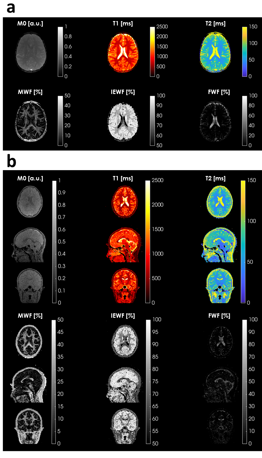
Figure 3 (a) 2D
MRF derived M0/T1/T2 and MWF/ IEWF/FWF maps of a representative subject. (b) 3D
MRF derived M0/T1/T2 and MWF/ IEWF/FWF maps of a representative subject (axial,
sagittal and coronal view).
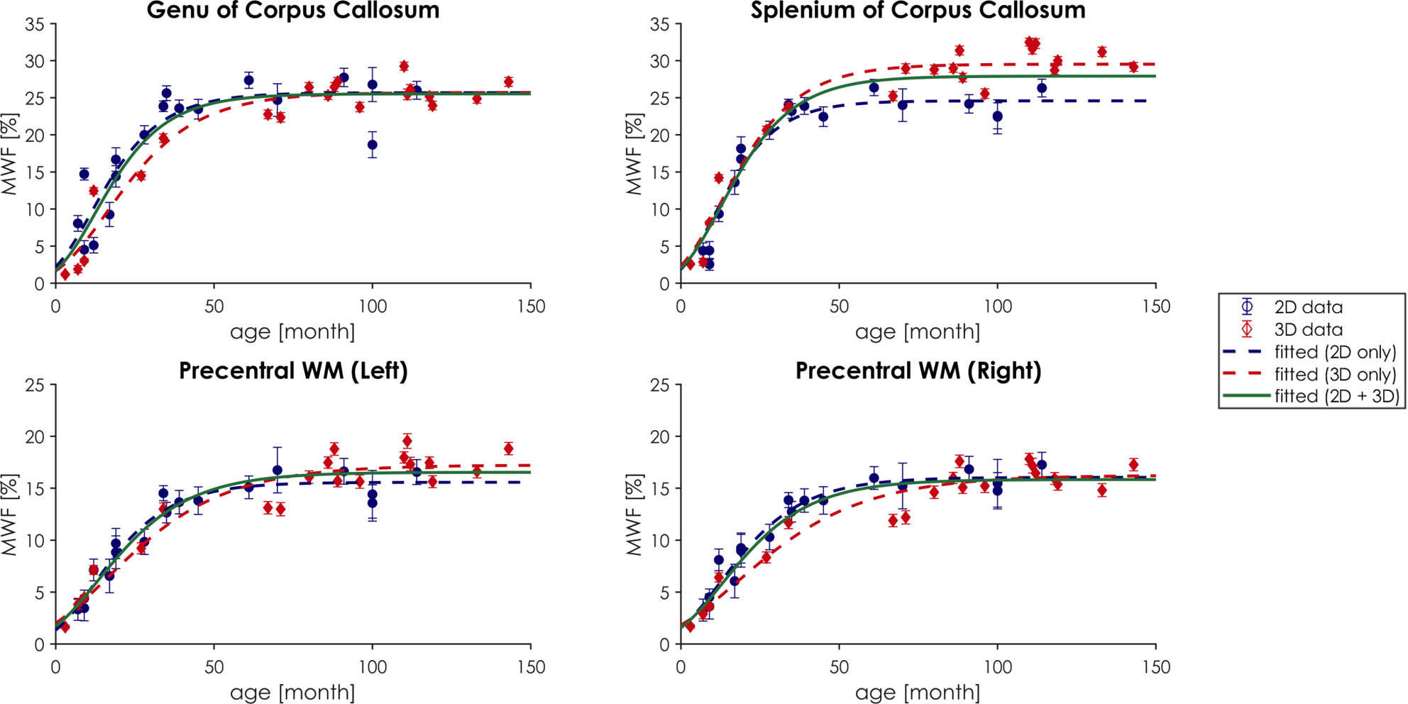
Figure 4 Myelin development curves in Genu
and Splenium of Corpus Callosum and Left/Right precentral White Matter.