Shohei Fujita1,2, Matteo Cencini3,4, Guido Buonincontri3,4, Naoyuki Takei5, Rolf F. Schulte6, Wataru Uchida1, Akifumi Hagiwara1, Koji Kamagata1, Osamu Abe2, Michela Tosetti3,4, and Shigeki Aoki1
1Department of Radiology, Juntendo University, Tokyo, Japan, 2Department of Radiology, The University of Tokyo, Tokyo, Japan, 3Imago7 Foundation, Pisa, Italy, 4IRCCS Stella Maris, Pisa, Italy, 5MR Applications and Workflow, GE Healthcare, Tokyo, Japan, 6GE Healthcare, Munich, Germany
1Department of Radiology, Juntendo University, Tokyo, Japan, 2Department of Radiology, The University of Tokyo, Tokyo, Japan, 3Imago7 Foundation, Pisa, Italy, 4IRCCS Stella Maris, Pisa, Italy, 5MR Applications and Workflow, GE Healthcare, Tokyo, Japan, 6GE Healthcare, Munich, Germany
The mean within-subject coefficients of variation of local thickness/volume, T1, and T2 of brain structures in healthy subjects undergoing three-dimensional MRF were 0.5–2.4% at 1.5 and 3T. Morphology was highly reproducible across field strengths.
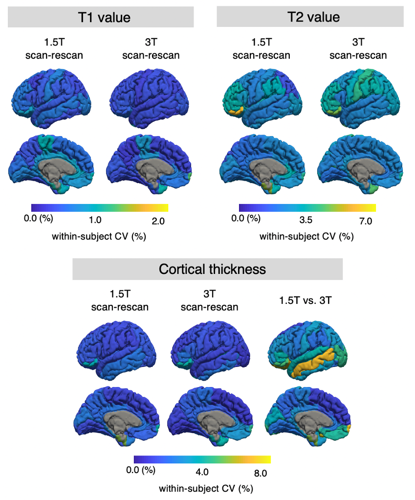
Figure 2. The scan-rescan within-subject coefficient of variance (wCV) of cortical thickness and relaxation times on cortical structures acquired with three-dimensional magnetic resonance fingerprinting. The scan-rescan wCVs of local cortical thickness, T1 value, and T2 value are overlaid on an inflated brain surface.
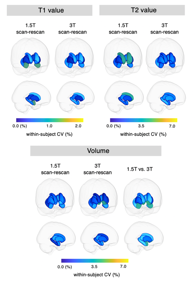
Figure 4. The scan-rescan within-subject coefficient of variance (wCV) of volume and relaxation times on subcortical structures acquired using three-dimensional magnetic resonance fingerprinting. The scan-rescan wCVs of subcortical volume, local T1 value, and local T2 value. Each subcortical structure is colored according to the regional wCV, and the brain surface is made transparent for the ease of visualization.