Thomas O`Reilly1, Peter Börnert1,2, Andrew Webb1, and Kirsten Koolstra1
1Radiology, Leiden University Medical Center, Leiden, Netherlands, 2Philips Research Hamburg, Hamburg, Germany
1Radiology, Leiden University Medical Center, Leiden, Netherlands, 2Philips Research Hamburg, Hamburg, Germany
In this work, we implemented a 3D MRF sequence at a 50 mT Halbach system to efficiently measure relaxation times in vivo.
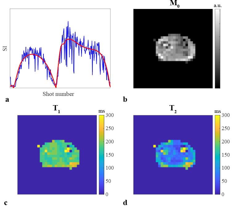
Figure 3. Matched MRF parameters
in a lower arm. (a) Measured signal curve (blue) and matched dictionary element
(red) in a voxel in the arm muscle. (b) Matched proton density map. (c) Matched
T1 map. (d) Matched T2 map. Parameter maps were set to
zero in the background.
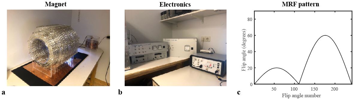
Figure 1. Experimental setup.
(a) 27 cm bore Halbach array with a 50 mT field at the center of the bore. (b)
Custom built gradient and RF power amplifiers are used to drive the gradient
and RF coils. The MRF sequence is run on a Magritek Kea2 spectrometer. (c) MRF
flip angle pattern of 240 pulses, preceded by an inversion pulse, used for the
MRF experiments.