Bernhard Gruber1,2, Jason P. Stockmann1, Azma Mareyam1, Boris Keil3, Anpreet Ghotra3, David A. Feinberg4, and Lawrence L. Wald1
1Department of Radiology, A.A. Martinos Center for Biomedical Imaging, Massachusetts General Hospital, Harvard Medical School, Charlestown, MA, United States, 2High Field MR Center, Center for Medical Physics and Biomedical Engineering, Medical University Vienna, Vienna, Austria, 3Department of Life Science Engineering, Institute of Medical Physics and Radiation Protection, Mittelhessen University of Applied Science, Giessen, Germany, 4Helen Wills Neuroscience Institute, University of California, Berkeley, Berkeley, CA, United States
1Department of Radiology, A.A. Martinos Center for Biomedical Imaging, Massachusetts General Hospital, Harvard Medical School, Charlestown, MA, United States, 2High Field MR Center, Center for Medical Physics and Biomedical Engineering, Medical University Vienna, Vienna, Austria, 3Department of Life Science Engineering, Institute of Medical Physics and Radiation Protection, Mittelhessen University of Applied Science, Giessen, Germany, 4Helen Wills Neuroscience Institute, University of California, Berkeley, Berkeley, CA, United States
We designed, built and simulated a 128-channel
receive array for cortical brain imaging at 7T to maximize SNR and achieve
highest accelerations needed for single-shot EPI based fMRI at sub-millimeter isotropic resolution.
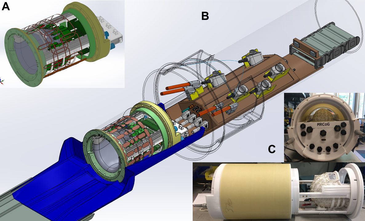
(A) 3D
rendering of the novel 128-ch brain array with a 24-ch
parallel Transmit Array setup. The pTx array is organized in 3 rows of 8. (B) 3D rendering of the whole 128-channel brain receive coil setup at 7 Tesla in SolidWorks
(Dassault Systems, France). The
table spoon extension (blue) with the Rx and Tx coils is connected to an interface box (brown) that slides into the MRI bore (C) Side
and back view of
the 128-ch coil array without populated elements. The
transmit (Tx) coils (1-ch birdcage or 24-ch pTx) slide over the receive (Rx)
array.
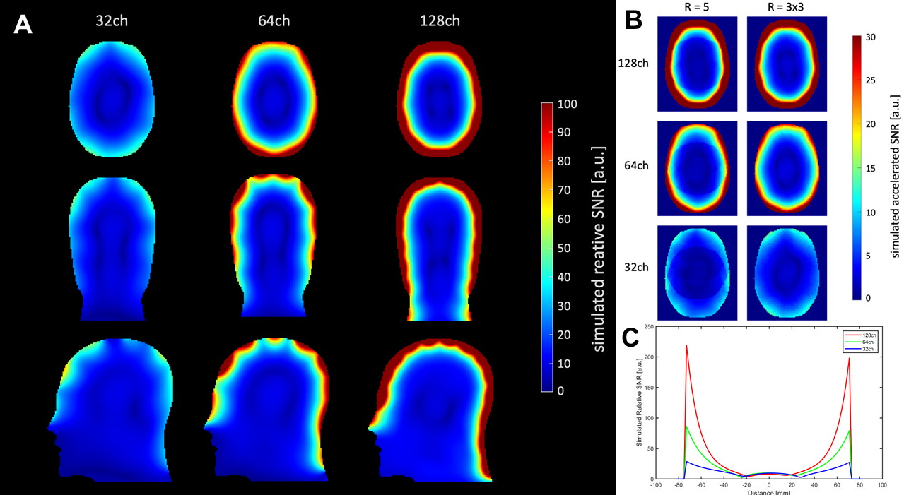
(A) Simulated
unaccelerated SNR maps of a 32-, 64-, and 128-channel array in a transverse,
coronal and sagittal slice. For simulation in MARIE a uniform head model (average
brain: epsilon=52, sigma=0.55 S/m), which closely matches the average brain
tissue, was used. (B) Simulated accelerated SNR maps for R=5 and R=3x3 of the
32-, 64-, and 128-channel array. (C) SNR profiles through a central axial slice
for the 128-channel (red line), 64-channel (green line), and 32-channel (blue
line).