Jiying Dai1,2, Tijl A. van der Velden1, Johannes M. Hoogduin1, Fabian Bartel2, Ettore F Meliadò1,2, Mark van Uden2, Catalina S. Arteaga de Castro2, Evita C. Wiegers1, Martijn Froeling1, Mark Gosselink1, Alexander J. E. Raaijmakers1,3, and Dennis W. J. Klomp1,4
1Radiology, UMC Utrecht, Utrecht, Netherlands, 2Tesla Dynamic Coils, Zaltbommel, Netherlands, 3Biomedical Engineering, Eindhoven University of Technology, Eindhoven, Netherlands, 4Tesla Engineering Ltd, West Sussex, United Kingdom
1Radiology, UMC Utrecht, Utrecht, Netherlands, 2Tesla Dynamic Coils, Zaltbommel, Netherlands, 3Biomedical Engineering, Eindhoven University of Technology, Eindhoven, Netherlands, 4Tesla Engineering Ltd, West Sussex, United Kingdom
With META Head coil, the acquisition time for 1H,
31P and 23Na in vivo scans is less than 25 minutes in
total, with high SNR, providing ample time to potentially add 13C
and/or 19F acquisitions in the same scan session following (labeled)
infusions.
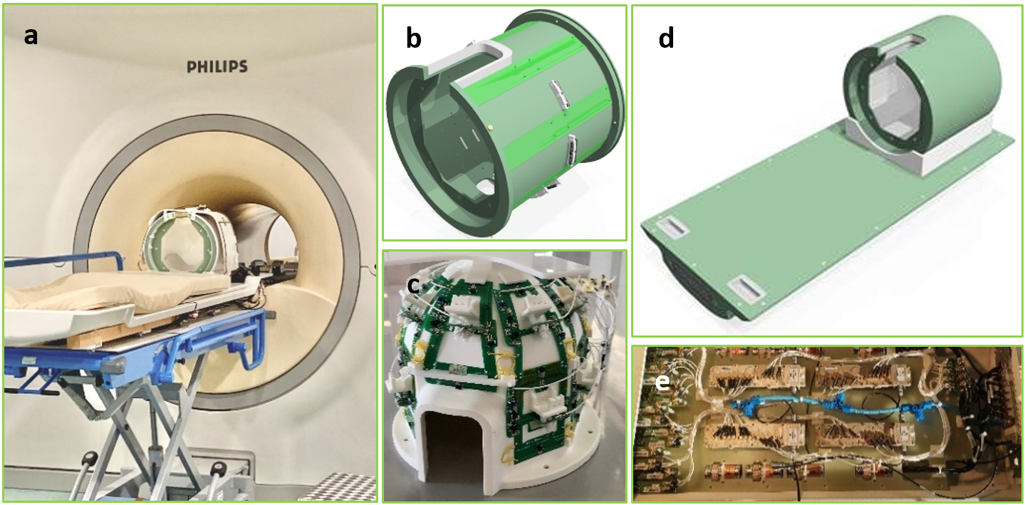
META Head Coil: a) The coil on the 7T MR system
including the 31P bore coil (embedded in bore) and the 23Na clamp; b) The eight-channel
dipole array as RF transceivers for 1H and 19F; c) The
fifteen-channel receiver array for 31P, 23Na and 13C;
d) The entire coil; e) The digital interface platform under the front cover of d.
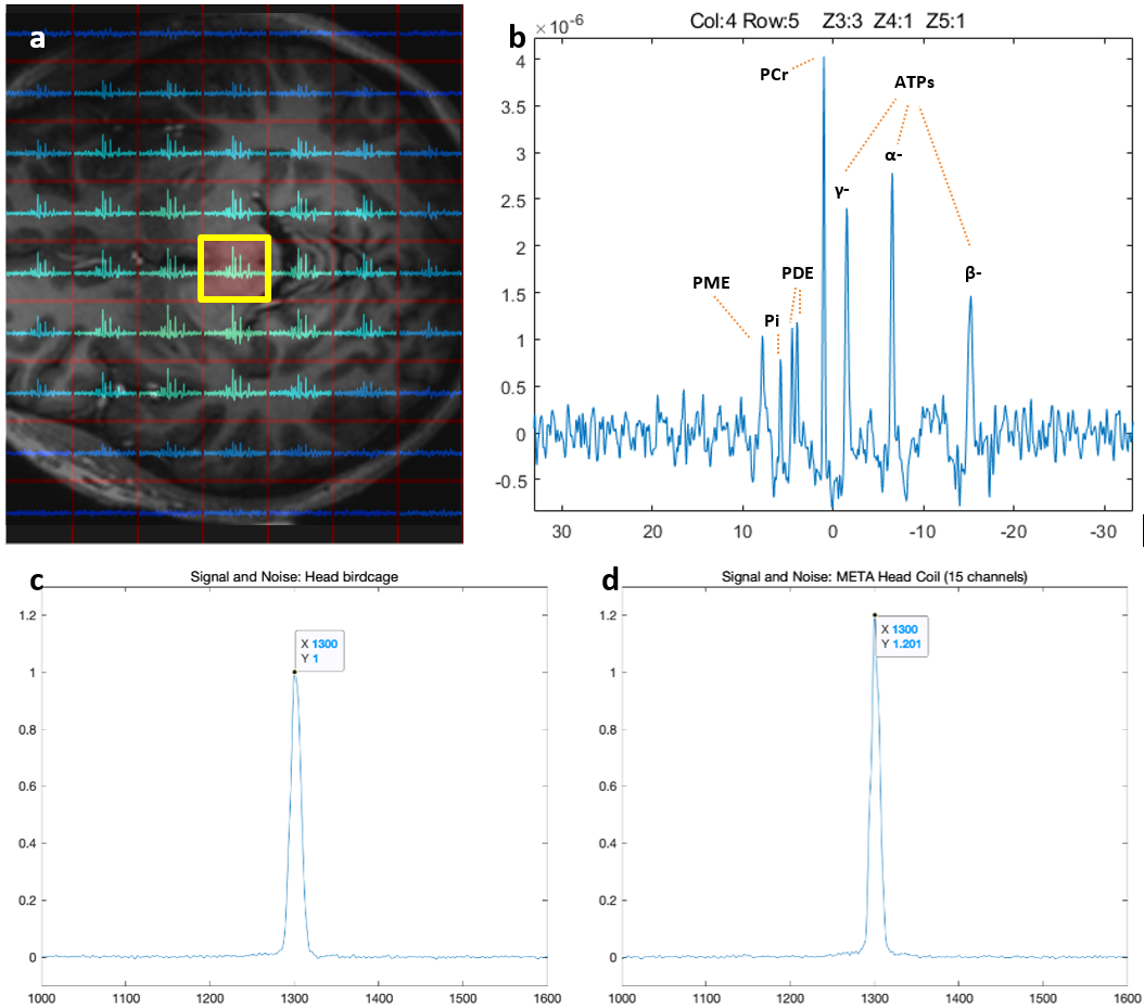
a) and b): Scan result from a brain 31P
CSI FID, TR=71.1ms, FA=13deg, voxel size =20*20*20mm3, Hamming-weighted NSA=70,
duration=10min: a) a slice from the 3D dataset showing the spectral map at full
brain coverage, the background image is overlaid manually for illustration; b) 31P spectrum
of the marked voxel of Figure a; c) and d): Signals from a close-fit head
birdcage[2] and the META head coil, at the center of a phantom. The
SNR comparison was assessed by scaling the data to obtain the same standard deviation
in the spectral noise then taking the integral of the signal in spectral domain.