Ibrahim A. Elabyad1, Maxim Terekhov1, and Laura M. Schreiber1
1Chair of Molecular and Cellular Imaging, Comprehensive Heart Failure Center (CHFC), University Hospital Wuerzburg, Wuerzburg, Germany
1Chair of Molecular and Cellular Imaging, Comprehensive Heart Failure Center (CHFC), University Hospital Wuerzburg, Wuerzburg, Germany
Parallel imaging with
acceleration factor R=6 was possible using Design2 while maintaining the mean g-factor
of 1.47 within the pig heart. Ultra-high-resolution
(0.35×0.35×4mm3) T2* weighted images were acquired on a fresh pig cadaver
using R=5.
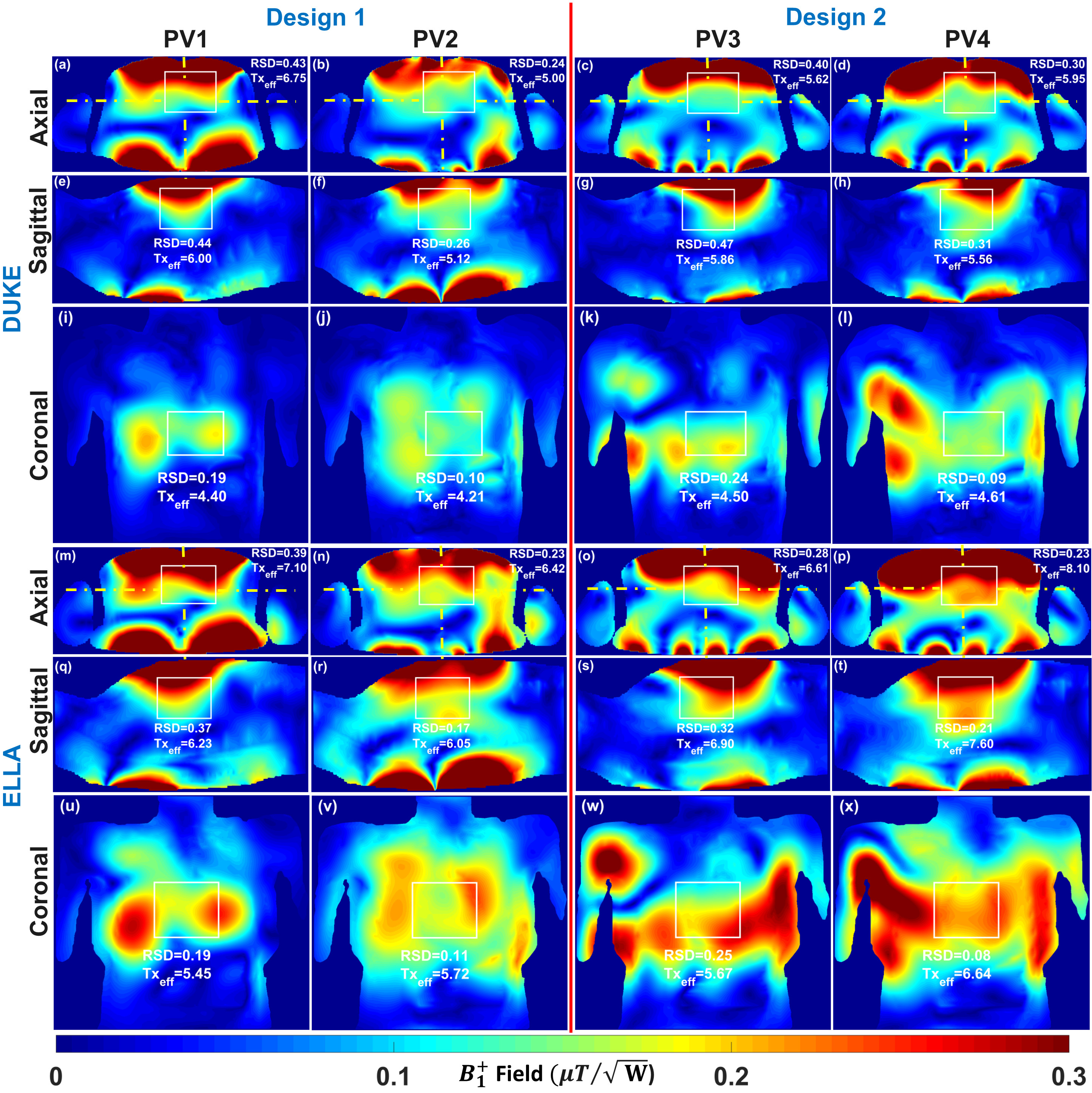
Figure
2. Simulated
central combined 2D $$${B_1^+}$$$-field distribution in central axial,
sagittal, and coronal planes
within Duke and Ella voxel models computed for the optimal phase vectors (PV1
and PV2 for Design1, and PV3 and PV4 for Design2). Both cardiac arrays were fed
with
and all coil
elements were fed with equal amplitudes. Values of RSD and mean $$$Tx_{eff}$$$ ($$$\mu{T}\sqrt{kW}$$$) were computed in the selected 2D ROI and written on
each image.
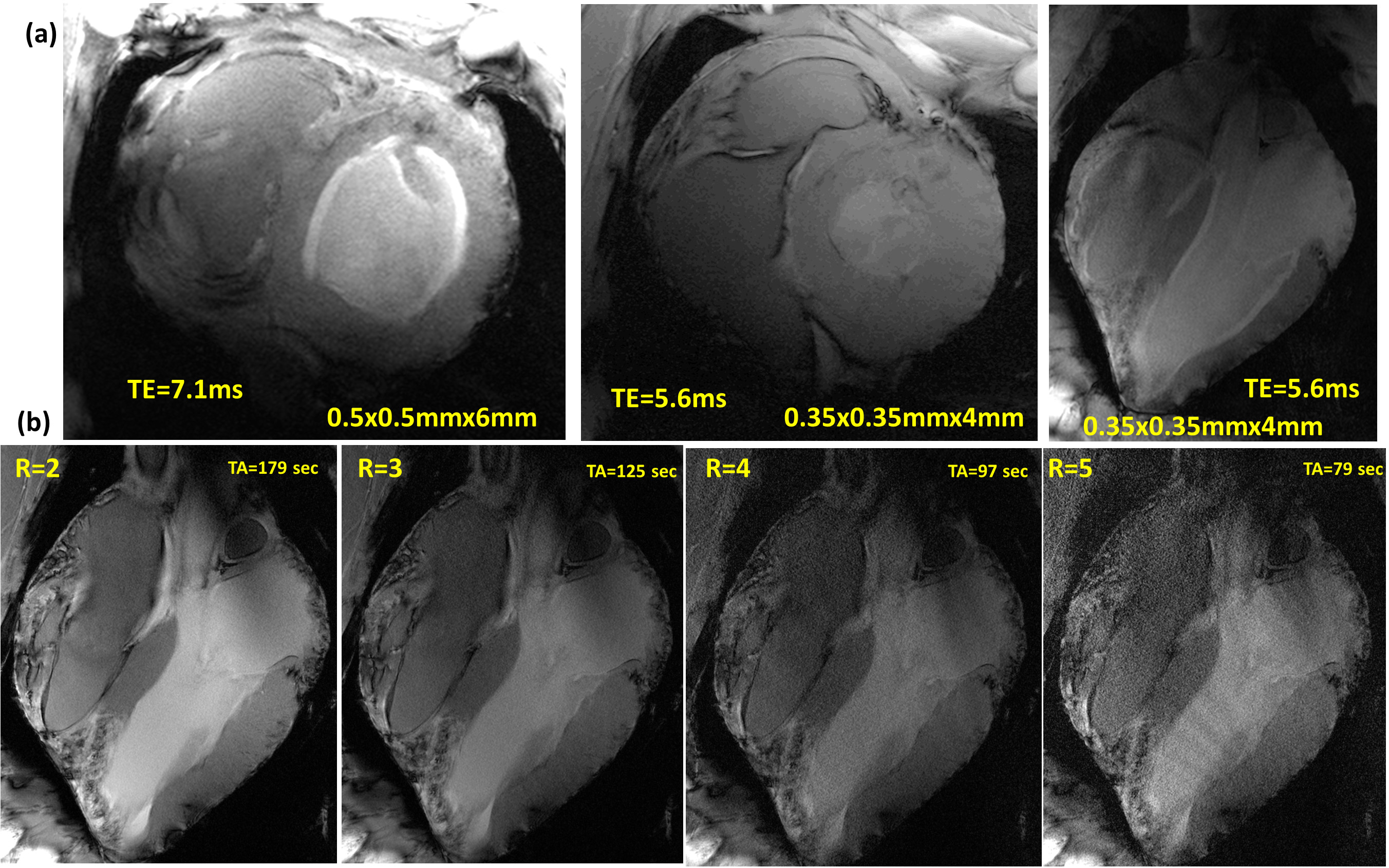
Figure 5. (a) Ultra-high
resolution T2* weighted images of the SA and LA heart views acquired with array
Design2. (b)
T2* weighted images of the LA view acquired with the spatial resolution
0.35×0.35×4mm3 at different GRAPPA acceleration factors to check parallel
imaging capability of the array Design2.