Guoxi Xie1, Hanwei Chen2, Chen Huang3, Xueping He2, Yueyuan Xie4, Xiaoyong Zhang5, Tianjing Zhang6, Yi Sun5, Debiao Li7, and Zhaoyang Fan8
1Department of Biomedical Engineering, Guangzhou Medical University, Guangzhou, China, 2Department of Radiology, Guangzhou Panyu Central Hospital, Guangzhou, China, 3Department of Minimally Invasive Interventional Radiology, Guangzhou Panyu Central Hospital, Guangzhou, China, 4Department of Anesthesiology, Mindong Hospital, Ningde, China, 5MR Collaborations, Siemens Healthcare Ltd, Shenzhen, China, 6Philips Healthcare, Guangzhou, China, 7Biomedical Imaging Research Institute, Cedars-Sinai Medical Center, Los Angeles, CA, United States, 8Department of Radiology, Keck School of Medicine, University of Southern California, Los Angeles, CA, United States
1Department of Biomedical Engineering, Guangzhou Medical University, Guangzhou, China, 2Department of Radiology, Guangzhou Panyu Central Hospital, Guangzhou, China, 3Department of Minimally Invasive Interventional Radiology, Guangzhou Panyu Central Hospital, Guangzhou, China, 4Department of Anesthesiology, Mindong Hospital, Ningde, China, 5MR Collaborations, Siemens Healthcare Ltd, Shenzhen, China, 6Philips Healthcare, Guangzhou, China, 7Biomedical Imaging Research Institute, Cedars-Sinai Medical Center, Los Angeles, CA, United States, 8Department of Radiology, Keck School of Medicine, University of Southern California, Los Angeles, CA, United States
The thrombus signal characteristics obtained on BTI imaging are valuable for assessing the prognosis of acute DVT and may aid in guiding the clinical treatment plan.
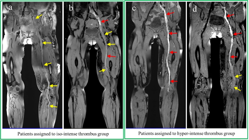
Figure 1. Representative black-blood
thrombus imaging (BTI) images from four patients who were categorized as
presenting with (a & b) iso- and (c & d) hyper- intense thrombus
signals, respectively. The iso-intense thrombi (yellow arrows) had
comparable signal intensity to the adjacent muscle while the hyper-intense
thrombi (red arrows) had higher signal intensity than the adjacent muscle. When
both iso- and hyper-intense thrombi were detected in a patient, the patient was
then assigned to the group that corresponded to the dominant thrombus signal (b
& d).
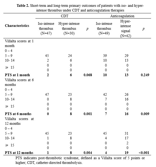
Table 2