John Heerfordt1,2, Aurélien Bustin2,3,4, Ludovica Romanin1,2, Estelle Tenisch2, Milan Prsa5, Tobias Rutz6, Christopher W. Roy2, Jérôme Yerly2,7, Juerg Schwitter6,8, Matthias Stuber2,7, and Davide Piccini1,2
1Advanced Clinical Imaging Technology, Siemens Healthcare AG, Lausanne, Switzerland, 2Department of Radiology, Lausanne University Hospital and University of Lausanne, Lausanne, Switzerland, 3IHU LIRYC, Electrophysiology and Heart Modeling Institute, Fondation Bordeaux Université, Pessac-Bordeaux, France, 4Department of Cardiovascular Imaging, Hôpital Cardiologique du Haut-Lévêque, CHU de Bordeaux, Pessac, France, 5Division of Pediatric Cardiology, Department Woman-Mother-Child, Lausanne University Hospital and University of Lausanne, Lausanne, Switzerland, 6Division of Cardiology, Cardiovascular Department, Lausanne University Hospital and University of Lausanne, Lausanne, Switzerland, 7CIBM Center for Biomedical Imaging, Lausanne, Switzerland, 8Cardiac MR Center, Lausanne University Hospital, Lausanne, Switzerland
1Advanced Clinical Imaging Technology, Siemens Healthcare AG, Lausanne, Switzerland, 2Department of Radiology, Lausanne University Hospital and University of Lausanne, Lausanne, Switzerland, 3IHU LIRYC, Electrophysiology and Heart Modeling Institute, Fondation Bordeaux Université, Pessac-Bordeaux, France, 4Department of Cardiovascular Imaging, Hôpital Cardiologique du Haut-Lévêque, CHU de Bordeaux, Pessac, France, 5Division of Pediatric Cardiology, Department Woman-Mother-Child, Lausanne University Hospital and University of Lausanne, Lausanne, Switzerland, 6Division of Cardiology, Cardiovascular Department, Lausanne University Hospital and University of Lausanne, Lausanne, Switzerland, 7CIBM Center for Biomedical Imaging, Lausanne, Switzerland, 8Cardiac MR Center, Lausanne University Hospital, Lausanne, Switzerland
Free-running
whole-heart MRI acquisitions can be sorted into motion-consistent clusters
prior to reconstruction based on the inherent similarities within the data. We showed that image quality
can be improved by enforcing total variation and/or low-rank constraints among clusters.
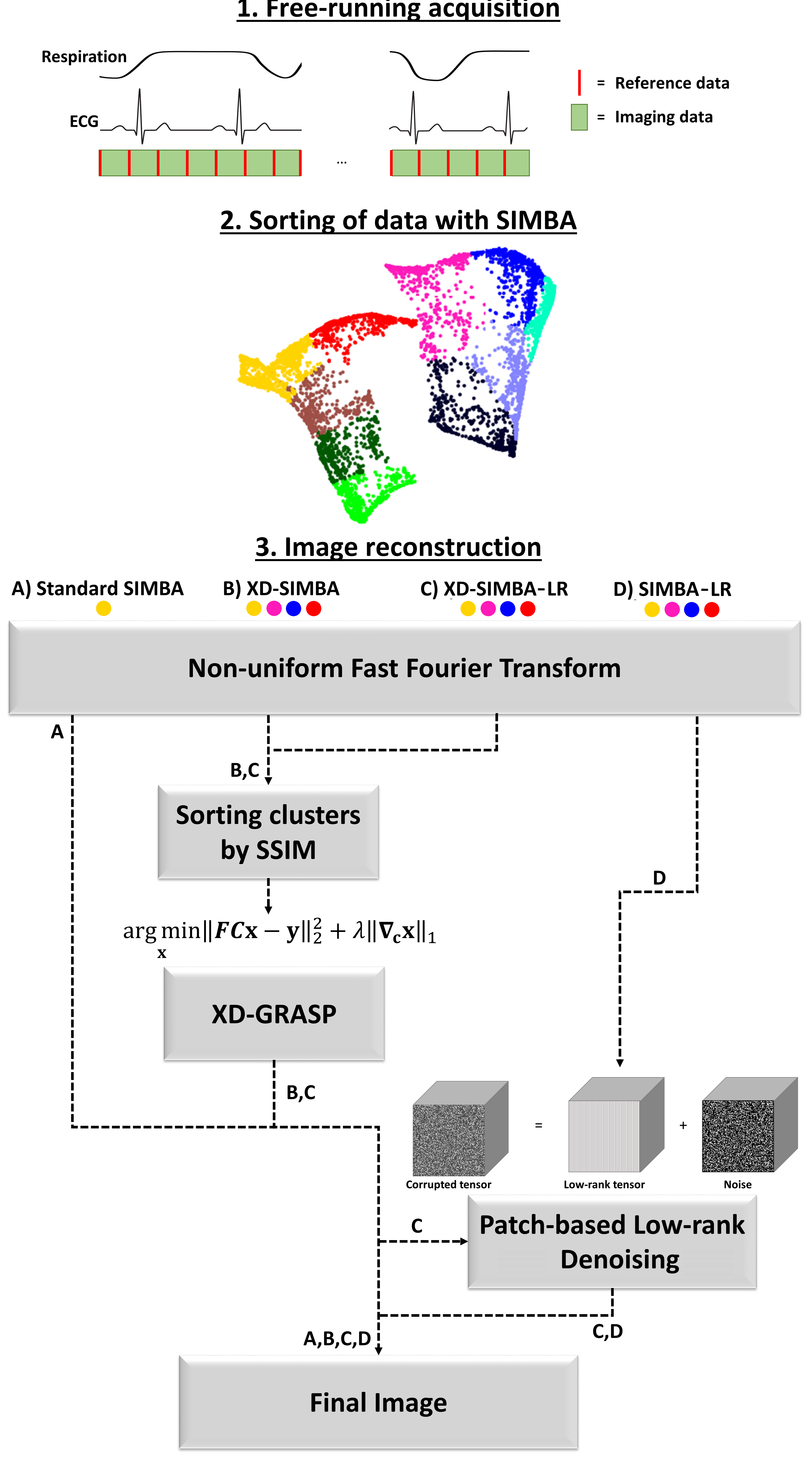
Figure 1. Overview of the different reconstructions compared in this work.
All reconstructions rely on the same SIMBA clustering of the
free-running data. A) Standard SIMBA consists of a non-uniform fast Fourier transform of the data in the most populated
cluster, B) XD-SIMBA consists of sorting the four most populated clusters based
on the SSIM prior to performing an
XD-GRASP reconstruction along the cluster
dimension, C) XD-SIMBA followed by patch-based low-rank denoising and D)
standard SIMBA followed by the same denoising algorithm applied to the four most populated clusters.
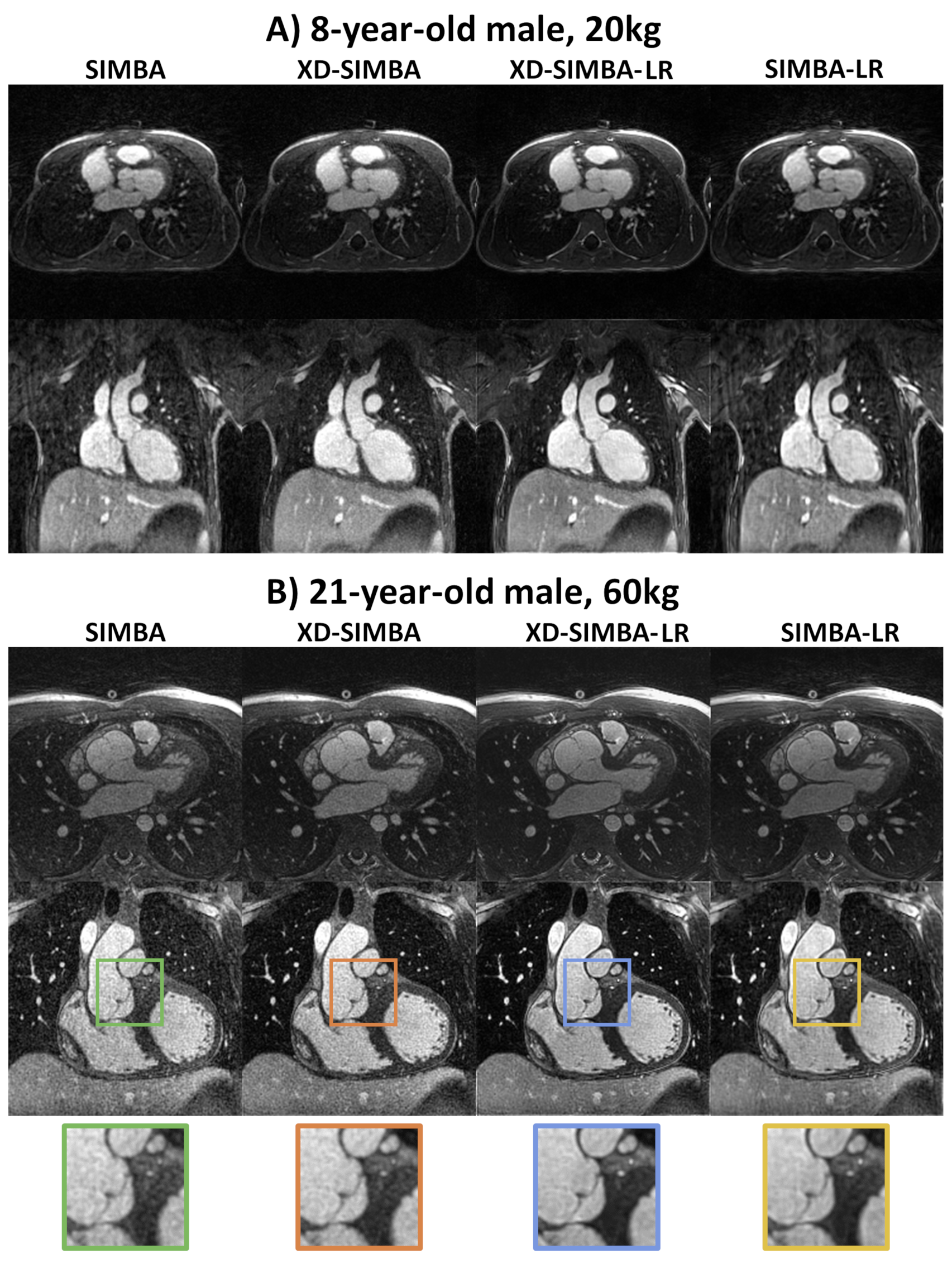
Figure 2. Representative image quality from one
pediatric and one adult patient.
A) The image characteristics differed between the various image
reconstruction methods. Methods that
share information between clusters generate less noisy images than the simple SIMBA reconstruction. B) In this adult patient, the aortic valve is depicted more clearly in
the XD-SIMBA-LR images compared to the other reconstruction types as seen in
the zoomed section. Finally, the cross-sections of the LAD and LCX coronary arteries appear
more conspicuous in the XD-SIMBA-LR approach.