Giulia MC Rossi1, Nemanja Masala1, Jessica AM Bastiaansen1, Aurelien Bustin1,2, Jérôme Yerly1,3, John Heerfordt1,4, Davide Piccini1,4, Matthias Stuber1,3, and Christopher W Roy1
1Department of Radiology, Lausanne University Hospital (CHUV) and University of Lausanne (UNIL), Lausanne, Switzerland, 2LIRYC (Electrophysiology and Heart Modeling Institute), Bordeaux, France, 3CIBM Center for Biomedical Imaging, Lausanne, Switzerland, 4Advanced Clinical Imaging Technology (ACIT), Siemens Healthcare AG, Lausanne, Switzerland
1Department of Radiology, Lausanne University Hospital (CHUV) and University of Lausanne (UNIL), Lausanne, Switzerland, 2LIRYC (Electrophysiology and Heart Modeling Institute), Bordeaux, France, 3CIBM Center for Biomedical Imaging, Lausanne, Switzerland, 4Advanced Clinical Imaging Technology (ACIT), Siemens Healthcare AG, Lausanne, Switzerland
The proposed auto-focusing method for free-running 3D
CMRA corrects for non-rigid respiratory motion and accounts for heart-rate
variability, providing comparable image quality to 5D imaging in significantly shorter
reconstruction times.
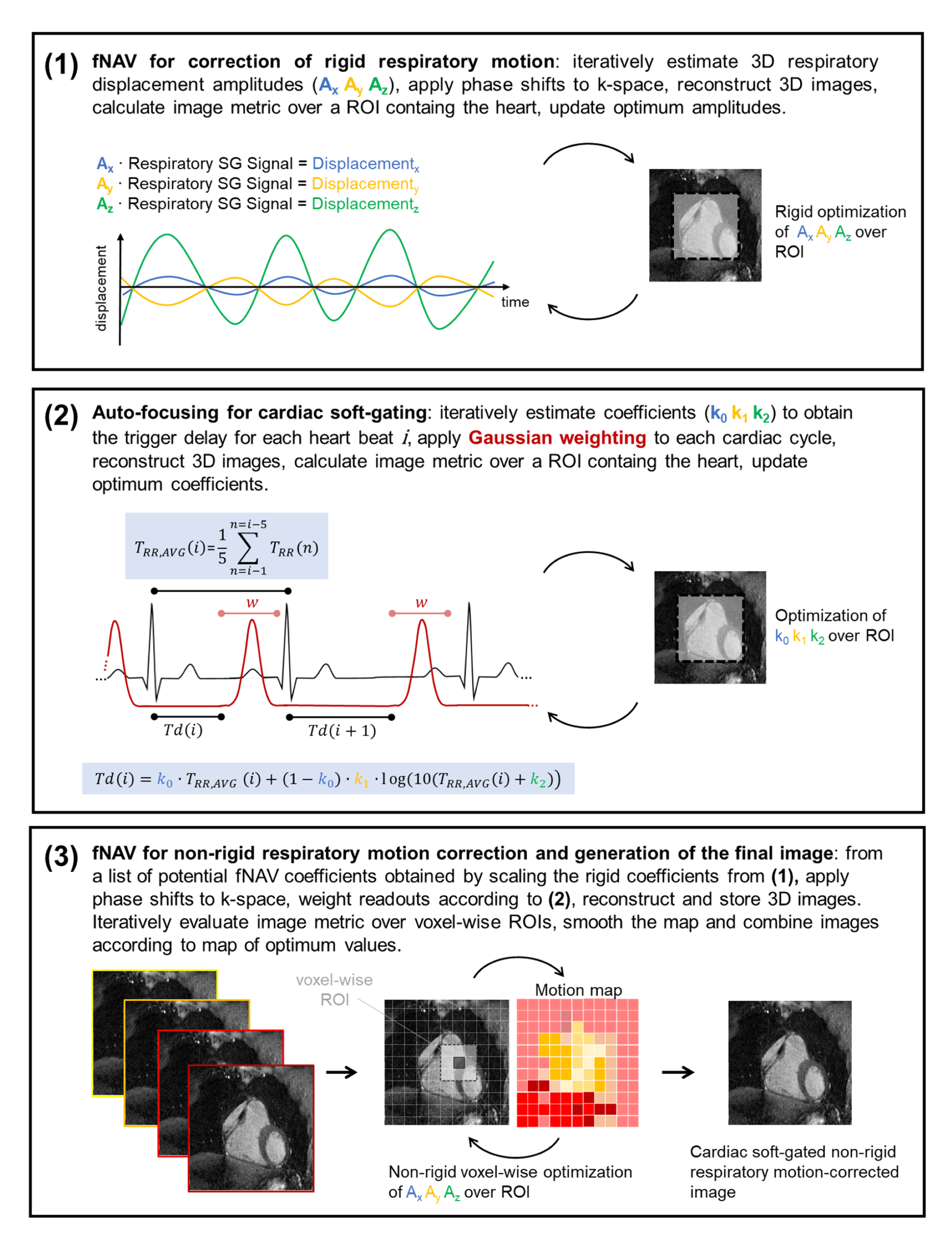
Figure
1. Overview of the 3-step auto-focusing framework for reconstructing cardiac
and respiratory motion-compensated 3D whole-heart CMRA images from free-running data.
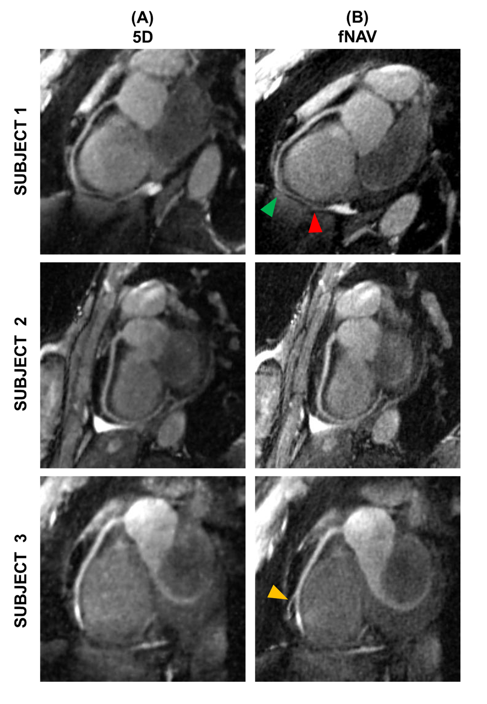
Figure 3.
Reformatted images of the right coronary artery (RCA). Reformatted images of the RCA obtained with Soap-Bubble
are shown for three representative volunteers for the reference 5D
reconstruction (A) and for the proposed fNAV-based reconstruction (B). Arrows
indicate RCA branches (yellow) or segments (green: beginning of the segment,
red: end of the segment) for which the fNAV-based reconstruction approach
allowed for improved detail visibility.