Ioannis Koktzoglou1,2, Rong Huang1, Nondas Leloudas1, and Robert R Edelman1,3
1Radiology, NorthShore University HealthSystem, Evanston, IL, United States, 2University of Chicago Pritzker School of Medicine, Chicago, IL, United States, 3Northwestern University Feinberg School of Medicine, Chicago, IL, United States
1Radiology, NorthShore University HealthSystem, Evanston, IL, United States, 2University of Chicago Pritzker School of Medicine, Chicago, IL, United States, 3Northwestern University Feinberg School of Medicine, Chicago, IL, United States
Leveraging multi-echo
stack-of-stars readouts and a novel inter-echo velocity extraction
computational framework, qTOF and qQISS MRA provide for simultaneous high-resolution
anatomic and quantitative hemodynamic evaluation of the intracranial arteries.
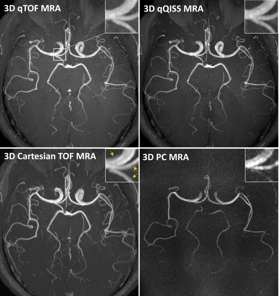
Figure 1. Transversal MIP
images of the brain showing the appearance of resolution-matched qTOF, qQISS, standard
Cartesian TOF, and 3D phase contrast (PC) MRA. Note the excellent correlation
of arterial anatomy of qTOF and qQISS with respect to standard TOF MRA, and the superior
SNR with respect to 3D PC MRA. qTOF and qQISS obtained using root-mean-square
combination of TE1 and TE3. Insets show that flow misregistration
artifact seen with Cartesian TOF MRA (arrows) is reduced with qTOF and qQISS.
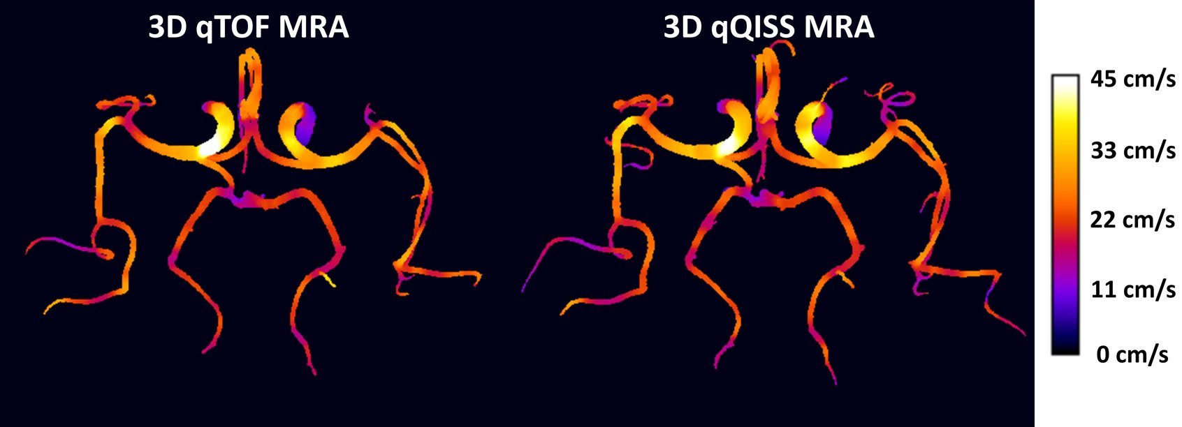
Figure 2. Transversal MIP
images showing mean cross-sectional velocity maps corresponding to the qTOF and
qQISS angiograms shown in Figure 1. Note the progressive reduction of blood flow
velocity from the larger to smaller arterial branches.