Chun-Hung Yeh1,2, Rung-Yu Tseng1, Susan Shur-Fen Gau3, and Hsiang-Yuan Lin4
1Institute for Radiological Research, Chang Gung University and Chang Gung Memorial Hospital, Taoyuan, Taiwan, 2Department of Child and Adolescent Psychiatry, Chang Gung Memorial Hospital, Linkou Medical Center, Taoyuan, Taiwan, 3Department of Psychiatry, National Taiwan University Hospital and College of Medicine, Taipei, Taiwan, 4Azrieli Adult Neurodevelopmental Centre and Adult Neurodevelopmental and Geriatric Psychiatry Division, Centre for Addiction and Mental Health, Department of Psychiatry, University of Toronto, Toronto, ON, Canada
1Institute for Radiological Research, Chang Gung University and Chang Gung Memorial Hospital, Taoyuan, Taiwan, 2Department of Child and Adolescent Psychiatry, Chang Gung Memorial Hospital, Linkou Medical Center, Taoyuan, Taiwan, 3Department of Psychiatry, National Taiwan University Hospital and College of Medicine, Taipei, Taiwan, 4Azrieli Adult Neurodevelopmental Centre and Adult Neurodevelopmental and Geriatric Psychiatry Division, Centre for Addiction and Mental Health, Department of Psychiatry, University of Toronto, Toronto, ON, Canada
Based on multi-band multi-shell diffusion MRI data and fixel-based analysis, reduced white matter fiber density in the corpus callosum associated with autism is driven by autistic individuals comorbid with developmental disabilities.
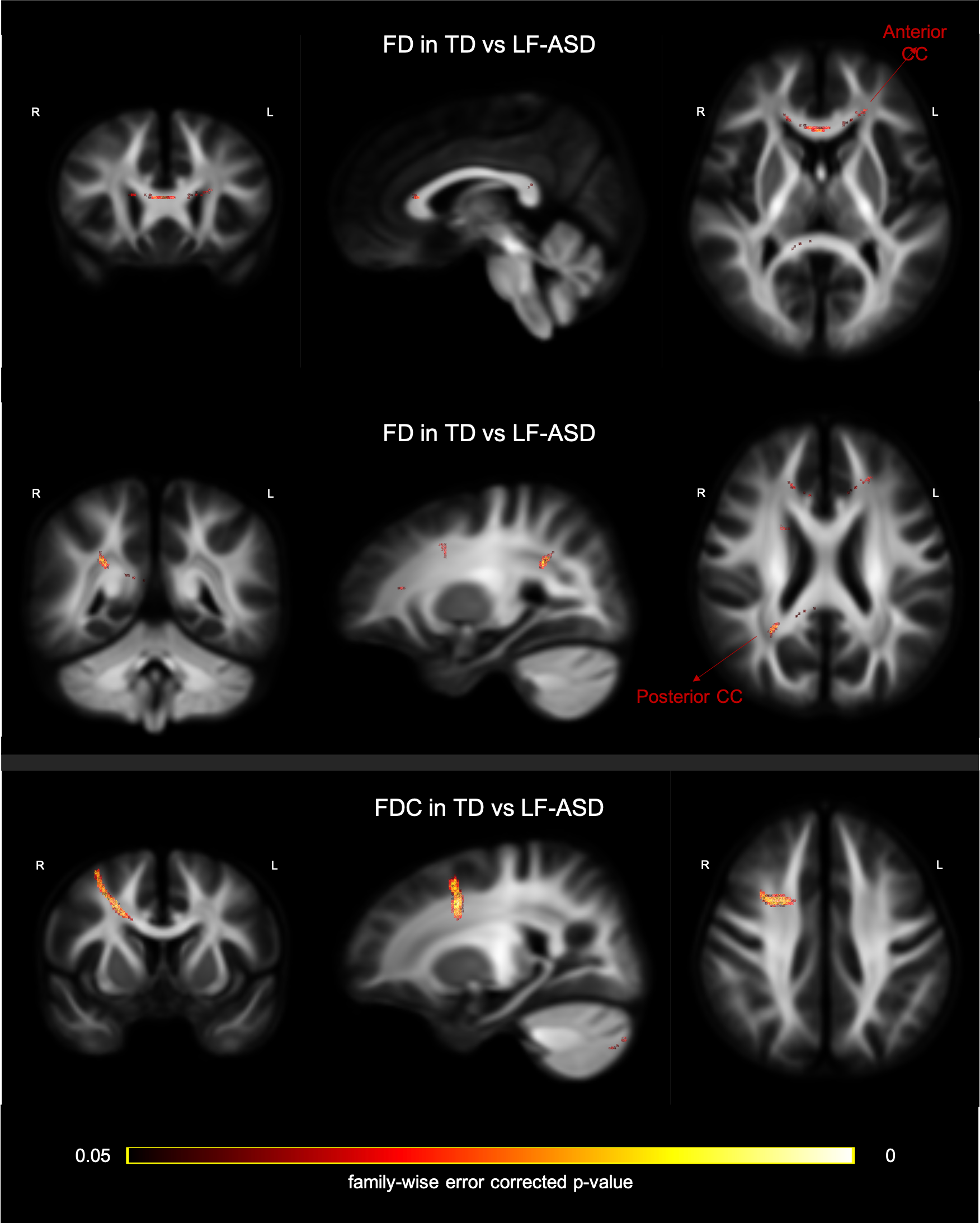
Figure 2: TD vs LF-ASD. Fixels are colour-coded by family-wise error corrected p-values and overlaid on the cohort FOD template. Upper and middle rows: Fixels with a significant (P<0.05) lower fibre density (FD) in the LF-ASD subgroup than TD group, illustrated at two slice locations per plane. Bottom row: Fixels with a significant (P<0.05) lower fibre density and cross-section (FDC) in the LF-ASD subgroup than TD group.
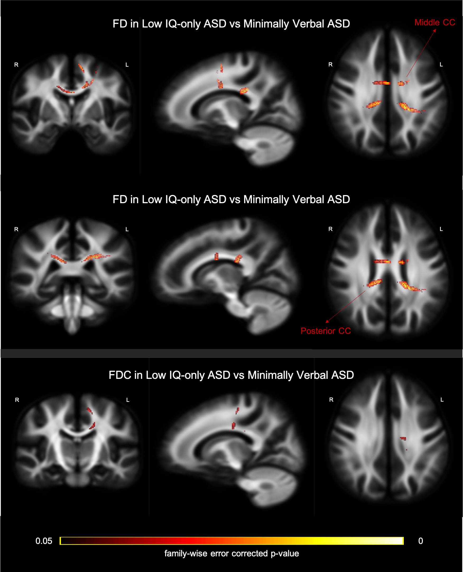
Figure 3: Low IQ ASD vs minimally verbal LF-ASD. Fixels are colour-coded by family-wise error corrected p-values and overlaid on the cohort FOD template. Upper and middle rows: Fixels with a significant (P<0.05) lower fibre density (FD) in the minimally verbal ASD than the low IQ-only subgroup, illustrated at two slice locations per plane. Bottom row: Fixels with a significant (P<0.05) lower fibre density and cross-section (FDC) in the minimally verbal ASD than the low IQ-only subgroup.