William S.H. Kim1,2, Mikaela K. Dimick3,4, Danielle Omrin4, Beverley A. Orser4,5, Benjamin I. Goldstein4,6, and Bradley J. MacIntosh1,2
1Department of Medical Biophysics, University of Toronto, Toronto, ON, Canada, 2Sunnybrook Research Institute, Toronto, ON, Canada, 3Department of Pharmacology and Toxicology, University of Toronto, Toronto, ON, Canada, 4Sunnybrook Health Sciences Centre, Toronto, ON, Canada, 5Department of Anesthesiology and Pain Medicine, University of Toronto, Toronto, ON, Canada, 6Department of Psychiatry, University of Toronto, Toronto, ON, Canada
1Department of Medical Biophysics, University of Toronto, Toronto, ON, Canada, 2Sunnybrook Research Institute, Toronto, ON, Canada, 3Department of Pharmacology and Toxicology, University of Toronto, Toronto, ON, Canada, 4Sunnybrook Health Sciences Centre, Toronto, ON, Canada, 5Department of Anesthesiology and Pain Medicine, University of Toronto, Toronto, ON, Canada, 6Department of Psychiatry, University of Toronto, Toronto, ON, Canada
Multiple post-label delay arterial spin labeling detected significant treatment effects on cerebral blood flow and arterial transit time change following nitrous oxide or midazolam treatment in a cohort of adults with treatment-resistant bipolar disorder.

Figure 2. Regional changes in CBF (left), ATT (middle), and aCBV (right). A) Mean CBF in the frontal, parietal, and temporal lobes. B) Mean ATT in frontal and parietal lobes. C) Mean aCBV in the bilateral insula. Dashed lines represent individual participants. Insets illustrate regions of interest.
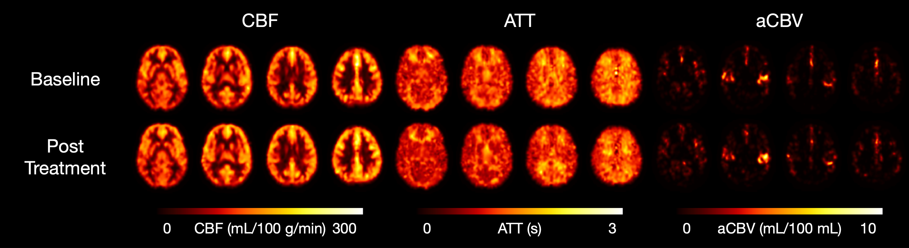
Figure 1. Representative CBF (left), ATT (middle), and aCBV (right) maps from a single participant in the nitrous oxide treatment arm at baseline (top row) and post-treatment (bottom row).
