Qiannan Zhao1, Hengyi Cao1,2,3, Yuan Xiao1, Qiyong Gong1, and Su Lui1
1Huaxi MR Research Center (HMRRC), Department of Radiology, West China Hospital, Sichuan University, Chengdu, China, 2Division of Psychiatry Research, Zucker Hillside Hospital, Glen Oaks, NY, United States, 3Center for Psychiatric Neuroscience, Feinstein Institute for Medical Research, Manhasset, NY, United States
1Huaxi MR Research Center (HMRRC), Department of Radiology, West China Hospital, Sichuan University, Chengdu, China, 2Division of Psychiatry Research, Zucker Hillside Hospital, Glen Oaks, NY, United States, 3Center for Psychiatric Neuroscience, Feinstein Institute for Medical Research, Manhasset, NY, United States
We identified two subgroups of patients with schizophrenia based on regional subcortical volume, displaying distinct degree of subcortical and global morphological alterations, and cognitive impairment.
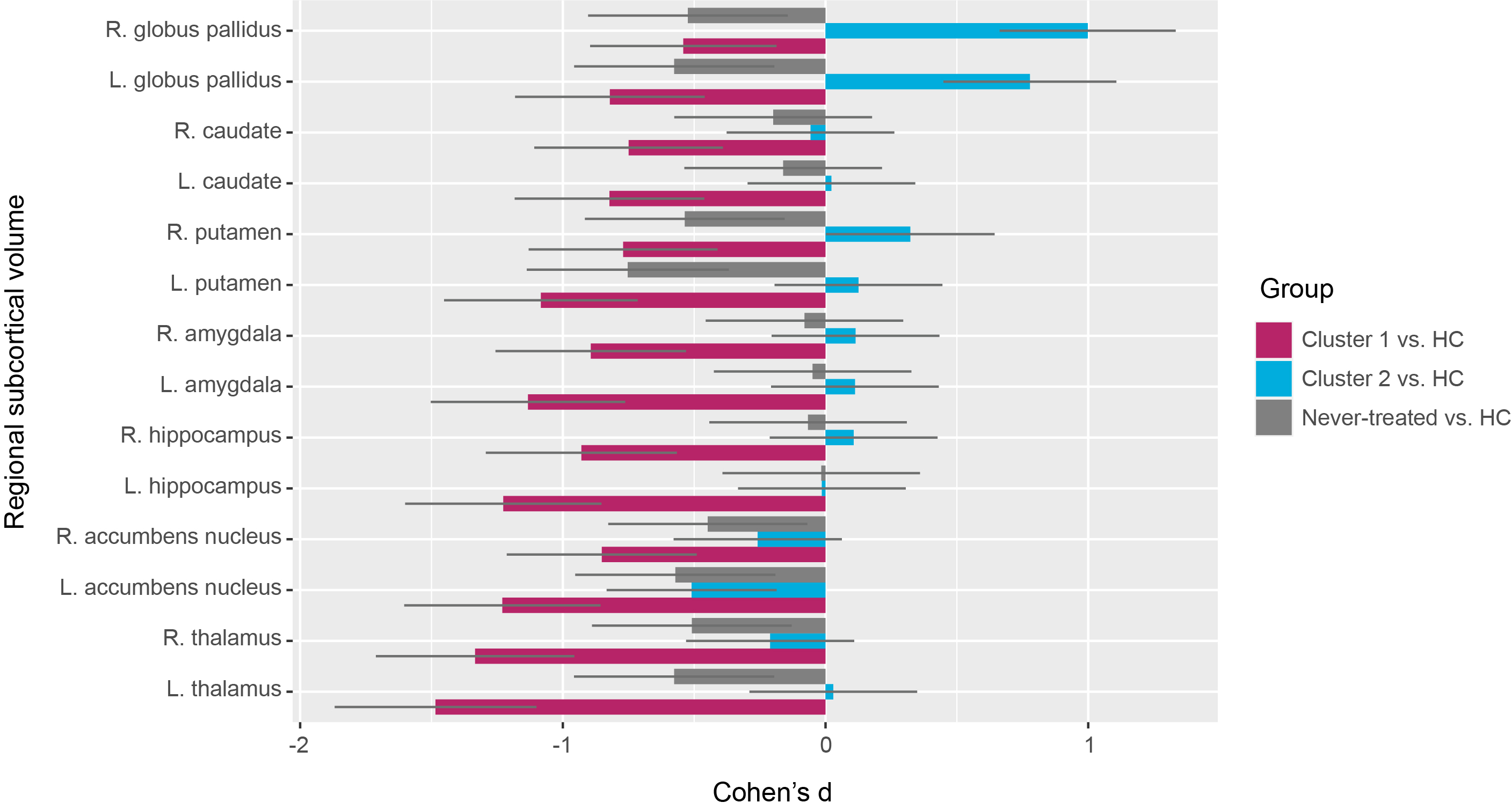
Fig. 1. Cohen’s d of regional
subcortical volume in two clusters of treated patients and the group of
never-treated patients compared with those in healthy controls. HC, healthy controls.
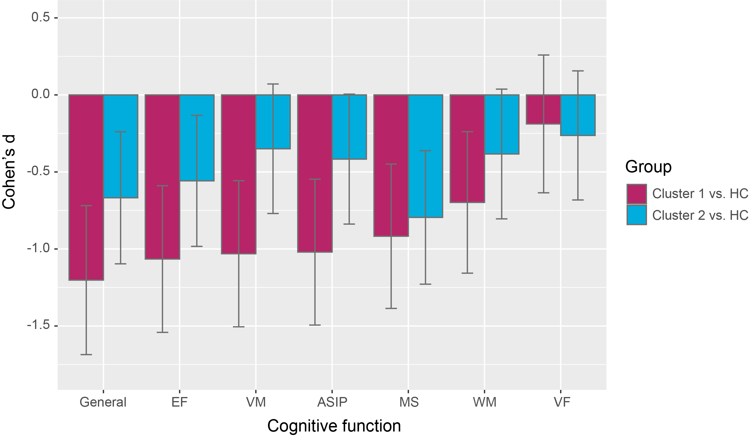
Fig. 2. Cohen’s d of cognitive function in two clusters of treated patients
compared with those in healthy controls. ASIP,
attention and speed of information processing; EF, executive functioning; HC, healthy
controls; MS, motor speed; VF, verbal fluency; VM, verbal memory; WM, working memory.