Lisa Novello1, Rafael Neto Henriques2, Andrada Ianuş2, Thorsten Feiweier3, Noam Shemesh2, and Jorge Jovicich1
1Center for Mind/Brain Sciences - CIMeC, University of Trento, Rovereto, Italy, 2Champalimaud Research, Champalimaud Centre for the Unknown, Lisbon, Portugal, 3Siemens Healthcare GmbH, Erlangen, Germany
1Center for Mind/Brain Sciences - CIMeC, University of Trento, Rovereto, Italy, 2Champalimaud Research, Champalimaud Centre for the Unknown, Lisbon, Portugal, 3Siemens Healthcare GmbH, Erlangen, Germany
We provide first evidence supporting the novel Correlation Tensor Imaging (CTI) framework to estimate non-Gaussian kurtosis sources in human brain tissue, in vivo, using a clinical 3T MRI system. Our findings augur well for future CTI applications.
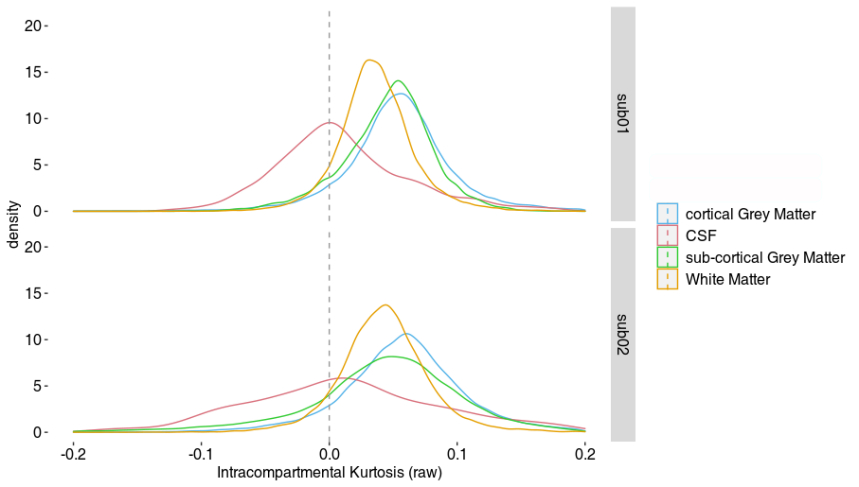
Figure 5: Human in vivo distributions of intra-compartmental kurtosis (KINTRA) in the tissues described in Fig. 4.
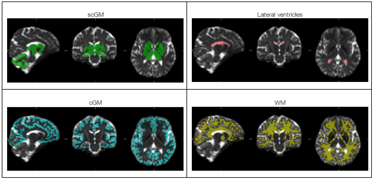
Figure 4: Representative tissue masks (subject 2) used to investigate intra-compartmental kurtosis distributions (Figure 5): subcortical gray matter (scGM), cortical GM (cGM), white matter (WM), and lateral ventricles cerebrospinal fluid (CSF).